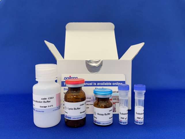
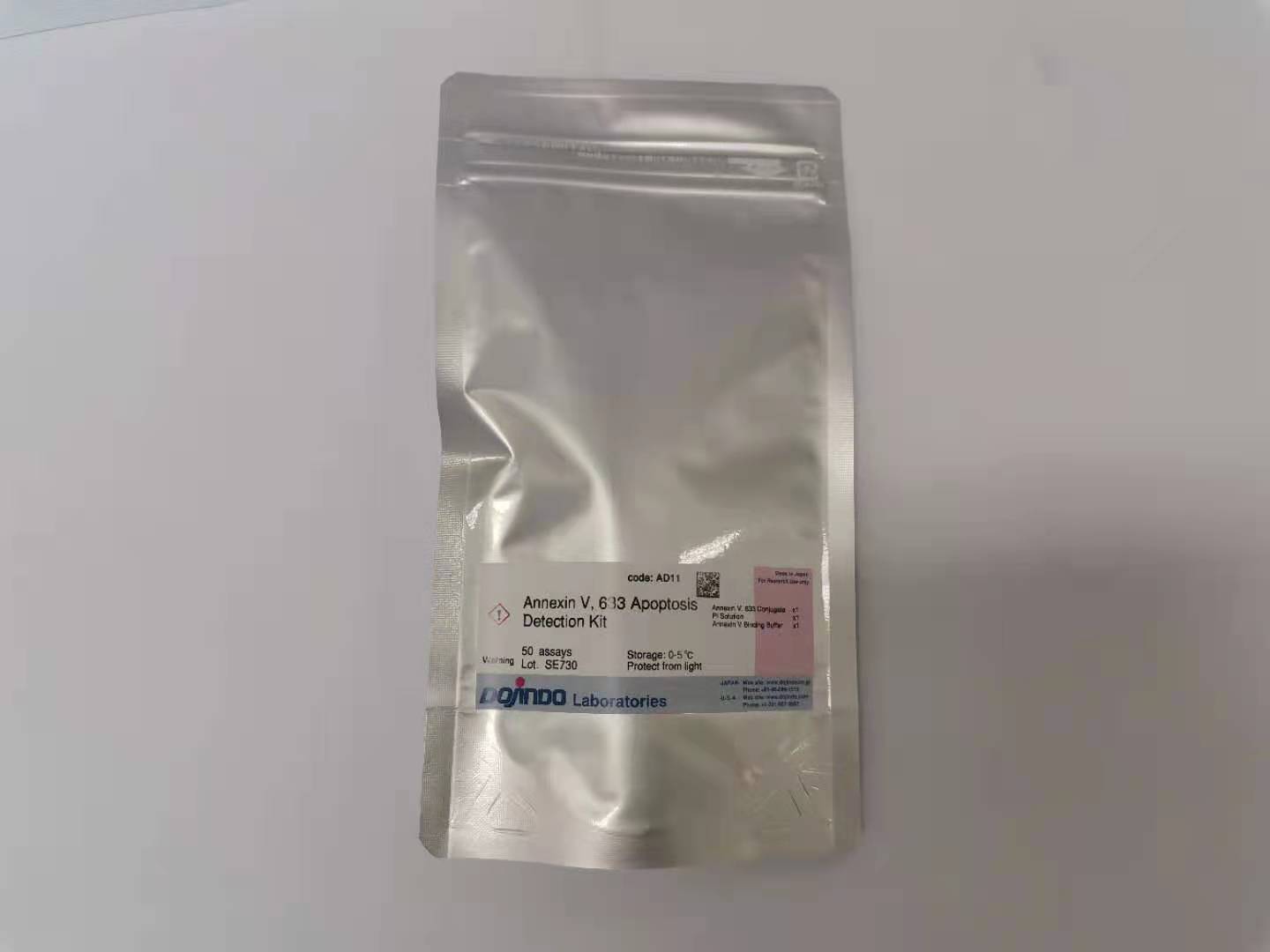
细胞凋亡检测试剂盒——Annexin V, 633 Apoptosis Detection Kit货号:AD11

细胞凋亡检测试剂盒——Annexin V, 633 Apoptosis Detection Kit货号:AD11
Annexin V, 633/PI凋亡检测试剂盒
Annexin V, 633 Apoptosis Detection Kit
商品信息
储存条件:0-5度保存,避光
运输条件:室温

特点:

· 灵敏度&稳定性高

· 性价比之选。

· 无需荧光补偿调节
下载说明书
产品文献
SDS下载
选择规格:
50assays
50assays*2
现货
细胞凋亡检测
活动进行中
试剂盒内含
产品概述
原理
荧光参数
注意事项
参考文献

活动进行中

订购满5000元,200元礼品等你拿

凑单关联产品TOP5

NO.1.    Cell Counting Kit-8    细胞增殖毒性检测

NO.2.    Annexin V, FITC Apoptosis Detection Kit    细胞凋亡检测

NO.3.    Cell Cycle Assay Kit    细胞周期检测

NO.4.    Caspase-3 Assay Kit-Colorimetric-    细胞凋亡检测

NO.5.    DAPGreen – Autophagy Detection   细胞自噬检测

试剂盒内含

1622170761260357.jpg

产品概述

细胞凋亡是指为维持有机体内环境稳定,由基因控制的细胞自主的有序的死亡。正常情况下任何细胞在形成过程中发生的异常都会通过凋亡消除。例如体内的癌细胞增长为肿瘤的过程会受细胞凋亡的引导而被抑制。然而在抑癌基因p53出现问题时,凋亡就不会诱导发生,从而导致癌细胞的不断增长。细胞凋亡可以通过细胞形态的变化或生物化学的变化来检测。目前常用的指标有caspase活性变化、DNA碎片、磷脂酰丝氨酸的外翻等。

原理

Annexin V染色的细胞可以用于检测细胞凋亡早期的细胞膜变化。在细胞凋亡早期,膜磷脂酰丝氨酸由脂膜内侧翻向外侧。Annexin V是一种分子量为35-36kD的Ca2+依赖性磷脂结合蛋白,与磷脂酰丝氨酸有高度亲和力,可通过细胞外侧暴露的磷脂酰丝氨酸与凋亡早期细胞的胞膜特异性结合,因此Annexin V 被作为检测细胞早期凋亡的灵敏指标之一。用红色荧光633标记的Annexin V通过流式细胞仪或荧光显微镜可以检测到细胞凋亡的发生。碘化丙啶 (Propidium Iodide, PI) 是一种核酸染料,PI只能透过凋亡晚期和死细胞的细胞膜,因此Annexin V和PI结合使用,可以区分凋亡早晚期的细胞及死细胞。

荧光参数

流式细胞仪检测

1. 加样到流式细胞仪检测,

633:激发波长Ex: 633 nm;

发射波长Em: 650-670 nm。

PI:激发波长Ex: 488 nm;

发射波长Em: 564-606 nm。

2. 633的红色荧光通过633通道(FL4)检测;

PI的红色荧光通过PI通道 (FL2或FL3)检测 (建议使用FL3)。

设定对照

1. 未染色细胞。

2. Annexin V, 633染色细胞 (没有PI)。

3. PI染色细胞 (没有Annexin V, 633)。

荧光显微镜检测

将细胞悬液滴到玻片上,置于显微镜上使用合适的滤光片检测。

* 由于EDTA会对细胞膜的特定部位有损伤,建议使用不含EDTA的胰酶消化贴壁细胞

注意事项

1. Annexin V, 633和PI均对光敏感。所有染色过程和培养过程均需避光。

2. 若细胞收集过程中使用胰酶,需设法去除残留的胰酶,这些胰酶会消化并降解Annexin V, 633,最终导致染色失败。

3. 用 APC和PE通道检测基本不用调节荧光补偿,但是如果细胞中还有其它荧光,例如细胞中有GFP,GFP荧光会影响PI通道的信息采集,需要调节荧光补偿。

参考文献

1. Deregulated lncRNA expression profile in the mouse lung adenocarcinomas with KRAS-G12D mutation and P53 knockout,Journal of Cellular and Molecular Medicine, 2019, 00:1-11

2. Curcumin Inhibits Growth of Human NCI-H292 Lung Squamous Cell Carcinoma Cells by Increasing FOXA2 Expression,Front. Pharmacol., 2018, doi: 10.3389/fphar.2018.00060

3. UCP2 ameliorates mitochondrial dysfunction, inflammation, and oxidative stress in lipopolysaccharide-induced acute kidney injury,International Immunopharmacology, 2019, 71, 336-349

4. γ-Glutamyl cyclotransferase contributes to endometrial carcinoma malignant progression and upregulation of PD-L1 expressionduring activation of epithelial-mesenchymal transition,International Immunopharmacology, 2019, 106039, doi:10.1016/j.intimp.2019.106039

关联产品
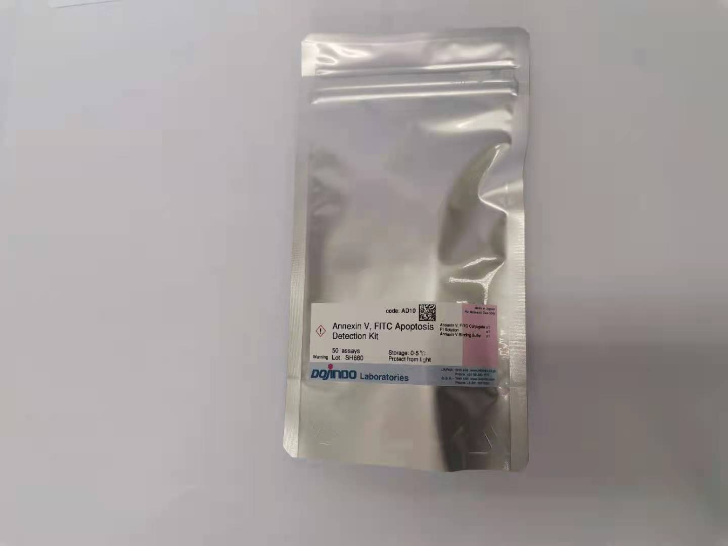
细胞凋亡检测试剂盒——Annexin V, FITC Apoptosis Detection Kit
Annexin V-FITC/PI凋亡检测试剂盒
Caspase-3 Assay Kit -Colorimetric-试剂盒
Caspase-3检测试剂盒-比色法

细胞凋亡检测试剂盒——Annexin V, FITC Apoptosis Detection Kit货号:AD10

细胞凋亡检测试剂盒——Annexin V, FITC Apoptosis Detection Kit货号:AD10
Annexin V-FITC/PI凋亡检测试剂盒
Annexin V, FITC Apoptosis Detection Kit
商品信息
储存条件:0-5度保存,避光
运输条件:室温

特点:

· 灵敏度&稳定性高

· 性价比高

· 荧光显微镜和流式检测双重方法检测

下载说明书
产品文献
SDS下载
选择规格:
50assays
50assays*2
价格:
细胞凋亡检测
活动进行中
试剂盒内含
产品概述
原理
荧光参数
相关资料
常见问题Q&A
参考文献

活动进行中

订购满5000元,200元礼品等你拿

凑单关联产品TOP5

NO.1.    Cell Counting Kit-8     细胞增殖毒性检测   

NO.2.    PI/RNase Staining    细胞周期检测   

NO.3.    Caspase-3 Assay Kit -Colorimetric    细胞凋亡检测  

NO.4.    ROS Assay Kit    活性氧检测

NO.5.    Calcein-AM/PI细胞双染试剂盒    活死细胞双染 

试剂盒内含

1622170154787265.jpg

产品概述

细胞凋亡是指为维持有机体内环境稳定,由基因控制的细胞自主的有序的死亡。正常情况下任何细胞在形成过程中发生的异常都会通过凋亡消除。例如体内的癌细胞增长为肿瘤的过程会受细胞凋亡的引导而被抑制。然而在抑癌基因p53出现问题时,凋亡就不会诱导发生,从而导致癌细胞的不断增长。细胞凋亡可以通过细胞形态的变化或生物化学的变化来检测。目前常用的指标有caspase活性变化、DNA碎片、磷脂酰丝氨酸的外翻等。

原理

Annexin V染色的细胞可以用于检测细胞凋亡早期的细胞膜变化。在细胞凋亡早期,膜磷脂酰丝氨酸由脂膜内侧翻向外侧。Annexin V是一种分子量为35-36kD的Ca2+依赖性磷脂结合蛋白,与磷脂酰丝氨酸有高度亲和力,可通过细胞外侧暴露的磷脂酰丝氨酸与凋亡早期细胞的胞膜特异性结合,因此Annexin V 被作为检测细胞早期凋亡的灵敏指标之一。用绿色荧光FITC标记的Annexin V通过流式细胞仪或荧光显微镜可以检测到细胞凋亡的发生。碘化丙啶 (Propidium Iodide, PI) 是一种核酸染料,PI只能透过凋亡晚期和死细胞的细胞膜,因此Annexin V和PI结合使用,可以区分凋亡早晚期的细胞及死细胞。

1622170486388694.jpg

Normal Cell                         Early stages of apoptosis                   Late stages of apoptosis

荧光参数

-流式细胞仪检测

1. 加样到流式细胞仪检测,激发波长Ex = 488 nm;发射波长Em = 530 nm。

2. Annexin V-FITC的绿色荧光通过FITC通道 (FL1) 检测;PI的红色荧光通过PI 通道 (FL2或FL3) 检测 (建议使用FL3)。

-荧光显微镜检测

将细胞悬液滴到玻片上,置于显微镜上使用合适的滤光片检测。* 由于EDTA会对细胞膜的特定部位有损伤,建议使用不含EDTA的胰酶消化贴壁细胞。

相关资料

所需的设备和材料

– 样品和诱导剂

– PBS、超纯水

– 合适量程的移液器

– 细胞培养用6,12,24,96孔板

– 流式细胞仪或荧光显微镜。

* 最大激发/发射波长  Annexin V, FITC: 494 nm/518 nm;PI: 535 nm/617 nm

溶液制备

1×Annexin V Binding solution:将10×Annexin V Binding Buffer用超纯水稀释10倍。

稳定性

试剂盒在0-5℃下可以保存6个月,请避光保存Annexin V, FITC结合物。

*实验实例及操作步骤请参考中文说明书

常见问题Q&A

Q1:贴壁细胞凋亡实验是否能够不消化?
A1:不选择消化的原因是什么?    =>主要是担心胰酶对细胞膜损伤而影响结果判断。

贴壁细胞在检测凋亡之前,如果没有出现团聚的情况,那么是可以尝试不消化直接镜检。理由是基于Annexin V- FITC与细胞反应的一个机理,凋亡早期的膜磷脂酰丝氨酸由膜内外翻,与Annexin V特异性结合,那么FITC标记的Annexin V呈现一个绿色荧光,所以我们保证足够的细胞膜暴露在工作液中,那即使没有经过胰酶消化,最终也是能够用荧光显微镜观察凋亡的情况。


不消化有几点好的地方:

1.避免一个对细胞膜的损伤,那结果能够很好的反应促凋亡的结果

2.操作简便,避免消化以及清洗过程中出现的操作失误


不消化有几点担心的地方:

1.无法制成细胞悬液,染色可能出现不充分

2.可能无法观察凋亡晚期PI和FITC双染的细胞

Q2:目前用Tunel的检测试剂盒做凋亡实验。和Tunel比较,用Annexin V-FITC的优点是什么?
A2: TUNEL这种方法灵敏度很高,但是特异性比较低,只能标记出凋亡中期和凋亡晚期的细胞,而且细胞坏死也会发生DNA断裂,不能算是真正的凋亡细胞,造成假阳性的现象。    建议老师两种方法都进行,以Tunel方法为主,Annexin V的方法为辅,这样两种方法都进行,互相也可以验证结果是否准确,互作参考。

参考文献

1. A Novel Allosteric Inhibitor of Phosphoglycerate Mutase 1 Suppresses Growth and Metastasis of Non-Small-Cell Lung Cancer,

Cell Metabolism, 2019, 30, 1-13

2. Tumor Microenvironment Activable Self-Assembled DNA Hybrids for pH and Redox Dual-Responsive Chemotherapy/PDT

Treatment of Hepatocellular Carcinoma, Advanced Science, 2017, 4(4), 1600460

3. Biogenic (R)-(+)-Lipoic Acid Only Constructed Cross-LinkedVesicles with Synergistic Anticancer Potency,

Advanced Functional Materials, 2018, 1806567

4. Anti-mitotic chemotherapeutics promote apoptosis through TL1A-activated death receptor 3 in cancer cells,

Cell Research, 2018, 28, 544-555

5. Bacteria-Driven Hypoxia Targeting for Combined Biotherapy and Photothermal Therapy, ACS Nano, 2018, 12(6), 5995-6005

6. Nanoenzyme-Augmented Cancer Sonodynamic Therapy by Catalytic Tumor Oxygenation, ACS Nano, 2018, 12, 3780-3795

7. Tumor-Specific Multiple Stimuli-Activated Dendrimeric Nanoassemblies with Metabolic Blockade Surmount Chemotherapy

Resistance, ACS Nano, 2017, 11(1), 416-429

8. Near Infrared-Guided Smart Nanocarriers for MicroRNA-Controlled Release of Doxorubicin/siRNA with Intracellular ATP as Fuel,

ACS Nano, 2016, 10(3), 3637-47

9. Noninvasive small-animal imaging of galectin-1 upregulation for predicting tumor resistance to radiotherapy,

Biomaterials, 2018, 158, 1-9

10. Cytotoxicity of guanine-based degradation products contributes to the antiproliferative activity of guanine-rich oligonucleotides,Chemical Science, 2015, 6, 3834-3836

11. A Novel Strategy through Combining iRGD Peptide with Tumor-icroenvironment-Responsive and Multistage Nanoparticles

for Deep Tumor Penetration, ACS Appl.Mater.Interfaces, 2015, 7(49), 27458-27466

12. JOSD1 inhibits mitochondrial apoptotic signalling to drive acquired chemoresistance in gynaecological cancer by stabilizing MCL1,Cell Death & Differentiation, 2020, 27, 55-70

13. The influence of printing parameters on cell survival rate and printability in microextrusion-based 3D cell printing technology,Biofabrication, 2015, 7(4), 045002

14. Downregulation of MCL-1 and upregulation of PUMA using mTOR inhibitors enhance antitumor efficacy of BH3 mimetics in triple-negative breast cancer, Cell Death & Disease, 2018, 9, 137

15. Mutations in the P10 region of procaspase-8 lead to chemotherapy resistance in acute myeloid leukemia by impairing procaspase-8 dimerization, Cell Death & Disease, 2018, 9(5), 516

16. Preclinical evaluation of antitumor of the proteasome inhibitor MLN2238 (ixazomib) in hepatocellular carcinoma cells,Cell Death & Disease, 2018, 9, 28

17. Long Noncoding RNA HCAL Facilitates the Growth and Metastasis of Hepatocellular Carcinoma by Acting as a ceRNA of LAPTM4B,Molecular Therapy: Nucleic Acids, 2017, 9, 440-451

18. TMEM74 promotes tumor cell survival by inducing autophagy via interactions with ATG16L1 and ATG9A,Cell Death & Disease, 2017, 8, e3031-3045

19. Nogo-C regulates cardiomyocyte apoptosis during mouse myocardial infarction, Cell Death & Disease, 2016, 7, e2432

20. TonEBP modulates the protective effect of taurine in ischemia-induced cytotoxicity in cardiomyocytes,Cell Death & Disease, 2015, 6, e2025

关联产品

Caspase-3 Assay Kit -Colorimetric-试剂盒
Caspase-3检测试剂盒-比色法
细胞凋亡检测试剂盒——Annexin V, 633 Apoptosis Detection Kit
Annexin V, 633/PI凋亡检测试剂盒
在线小工具

实验工具|稀释计算器|摩尔浓度计算器