上海金畔生物代理Hampton research品牌蛋白结晶试剂耗材工具等,我们将竭诚为您服务,欢迎访问Hampton research官网或者咨询我们获取更多相关Hampton research品牌产品信息。Products > 96 Well Crystallization Plates > Paul Marienfeld > LCP Sandwich Set
LCP Sandwich Set
Applications
- Crystallization screening of membrane proteins in lipidic mesophase as well as bicelle method and batch method
Features
- Small volume LCP, bicelle and microbatch crystallization
- High quality glass optics
- Excellent drop optics since mesophase bolus is physically sandwiched between two optically clear surfaces eliminating the mesophase / aqueous medium interface and the corresponding roughness
- Glass plate allows for visualization of microcrystals using birefringence-free examination between crossed polarizers
- Superhydrophobic glass surfaces
- Optimized for bright field, UV and fluorescent microscopy
- Excellent stability, resistant to evaporation
Description
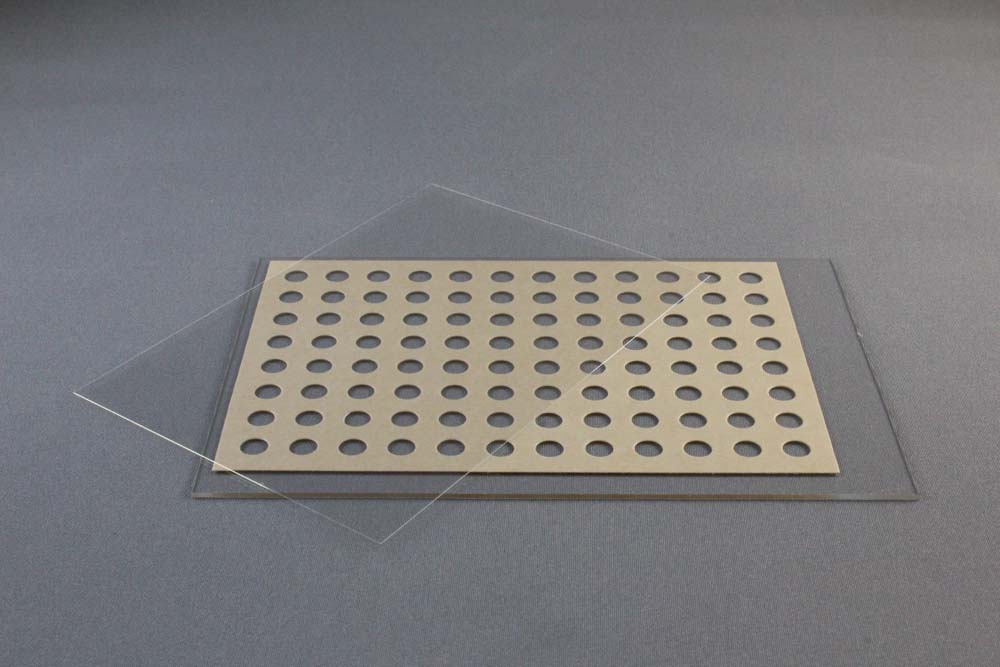
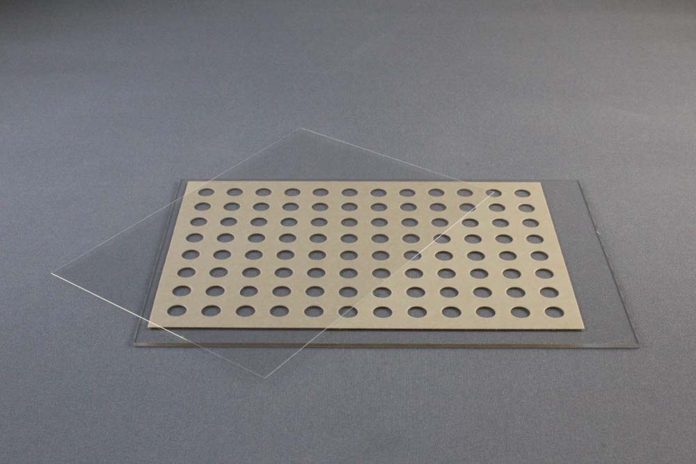
The LCP Sandwich Set consists of a base glass slide and an optimized cover slip. LCP Sandwich Set developed jointly with the renowned Scripps Research Institute in La Jolla, California, USA and is manufactured by Paul Marienfeld (0890003).
The LCP Glass Sandwich Set is a specially designed plate for either automated or manual setting of 96 Lipidic Cubic Phase matrix screening experiments. The plate can also be used for the bicelle method and batch method. The thin (< 2 mm high) plates have exquisite optical properties and are well suited for the detection of microcrystals and for birefringence-free imaging between crossed polarizers. The plates allow for in meso crystallization trials with 50 nanoliters protein/lipid mesophase and 1 microliter precipitant solution per trial.
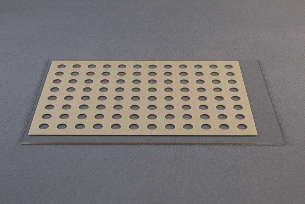
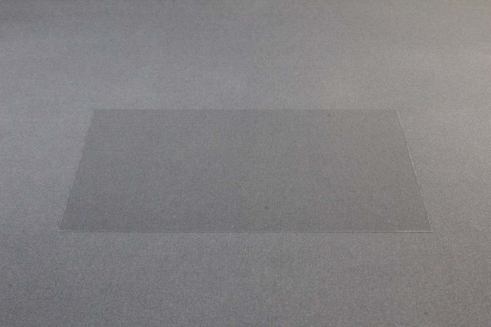
The 96 well LCP Glass Sandwich Plate consists of a) 127.8 x 85.5 x 1 mm glass base plate with the footprint of an SBS-compliant plate, a 140 µm thick double sticky spacer with 96 punched out holes and b) a 0.2 mm thick glass coverslip. The double sticky spacer is already adhered to the base plate. A brown paper liner covers and protects the top of the double sticky spacer and base plate. The 0.2 mm thick glass coverslip fits over and seals the entire 96 well lower plate. Alternatively a series of twenty-four siliconized 18 x 18 mm No. 1 square cover slides (HR3-152) can be used to seal the entire 96 well lower plate. Each 18 x 18 mm No. 1 cover slide seals 4 wells. Space for a bar code is available at the left end of the plate. Each well can contain 50 nanoliters of cubic phase and 1 microliter of crystallization reagent.
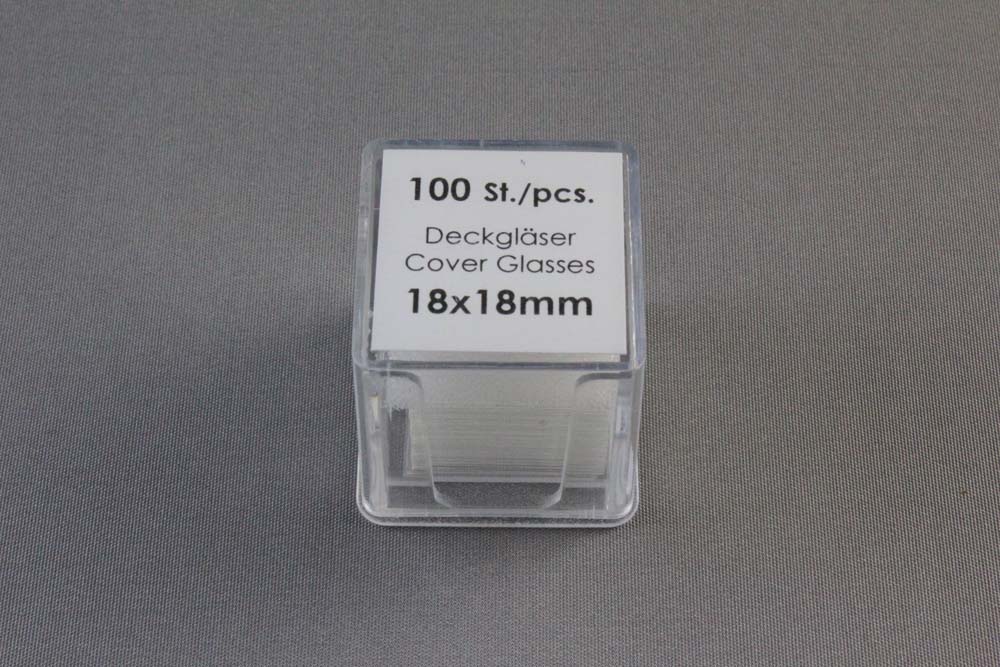
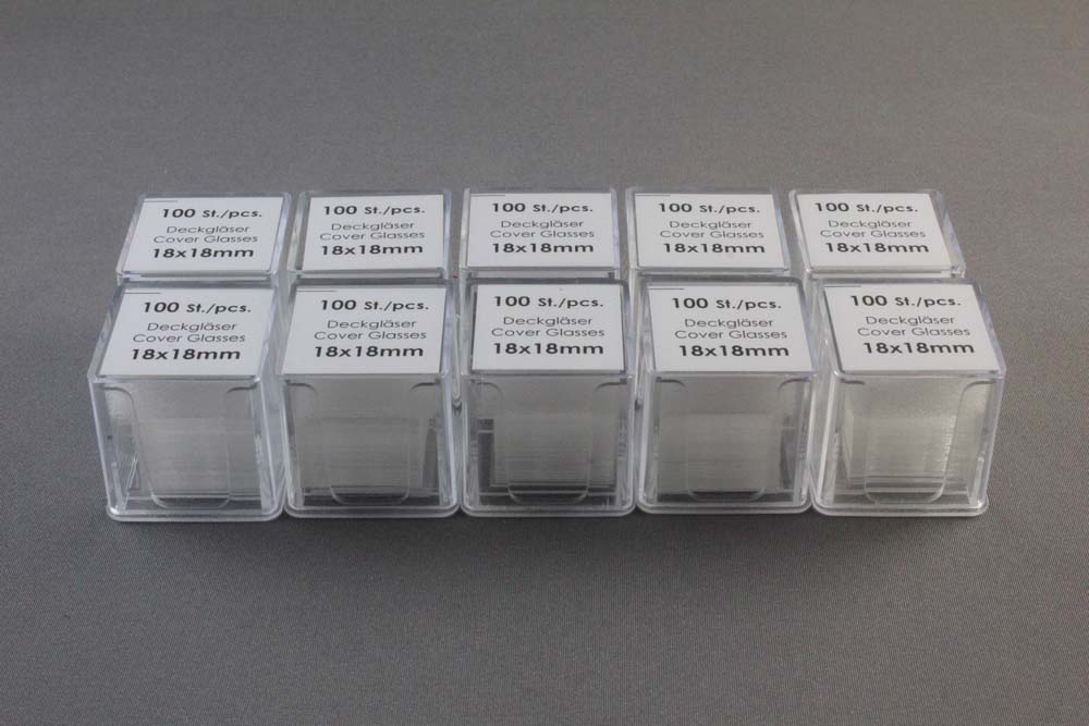
18 x 18 mm No. 1 Siliconized Cover Slides are made from chemically resistant borosilicate glass D 263 M of the first hydrolytic class. The slides are colorless, clear, and suitable for fluorescence microscopy. The slides feature a super hydrophobic surface on both sides. The slides are No. thickness (0.13 ro 0.16 mm). The slides are supplied in a two compartment plastic box, 100 slides per box, 10 boxes per case, 1,000 slides total for HR3-152.
The Tungsten-carbide glass cutter can be used for cutting and removal of the upper coverglass that seals the LCP Sandwich Set. Sold separately.
CAT NO
HR3-151
NAME
DESCRIPTION
Pack of 20
PRICE
$458.00
cart quote
CAT NO
HR3-152
NAME
DESCRIPTION
Case of 10 packs
PRICE
$322.00
cart quote
Support Material(s)
 HR3-151 LCP Sandwich Set User Guide
 HR3-151 LCP Sandwich Set Dimension Drawing
 Related Item(S)
- 4 Inch Soft Rubber Brayer
- Tungsten Carbide Scribe & Glass Cutter with Replaceable Tips
References
1. Nano-volume plates with excellent optical properties for fast, inexpensive crystallization screening of membrane proteins. Vadim Cherezov and Martin Caffrey. J. Appl. Cryst. (2003). 36, 1372-1377.
2. A robotic system for crystallizing membrane and soluble proteins in lipidic mesophases. Vadim Cherezov, Avinash Peddi, Lalitha Muthusubramaniam, Yuan F. Zheng and Martin Caffrey. Acta Cryst. (2004). D60, 1795-1807 DOI: 10.1107/S0907444904019109.
3. Bicelle crystallization: a new method for crystallizing membrane proteins yields a monomeric bacteriorhodopsin structure. Faham, S. & Bowie, J. U. (2002). J. Mol. Biol. 316, 1-6.


