细胞活性/毒性双重检测试剂盒——Viability/Cytotoxicity Multiplex Assay Kit货号:CK17

特色

产品中心细胞增殖/毒性细胞活性/毒性双重检测试剂盒——Viability/Cytotoxicity Multiplex Assay Kit
细胞活性/毒性双重检测试剂盒——Viability/Cytotoxicity Multiplex Assay Kit货号:CK17
细胞活性/毒性双重检测试剂盒
Viability/Cytotoxicity Multiplex Assay Kit
商品信息
储存条件:0-5度保存
运输条件:室温

特点:

● CCK-8与LDH双指标检测毒性,数据更全面。

● 准备1次细胞,同时检测2项数据。

● 内含500T的CCK-8与LDH检测试剂盒,

价格优于单独购买。

下载说明书
SDS下载
选择规格:
500 tests
现货
双重实验同时检测
性价比高
CCK-8&LDH
更高灵敏度检测方法CCK-L(点击查看)
活动进行中
试剂盒内含
产品概述
产品特点
检测原理
同时检测的操作方法
性价比高
参考文献

活动进行中

订购满5000元,200元礼品等你拿

凑单关联产品TOP5

NO.1.    ROS Assay Kit -Highly Sensitive DCFH-DA-    ROS检测

NO.2.    Annexin V, FITC Apoptosis Detection Kit    细胞凋亡检测

NO.3.    Calcein-AM/PI Double Staining Kit    活死细胞双染检测

NO.4.    Cell Cycle Assay Kit    细胞周期检测

NO.5.    Caspase-3 Assay Kit-Colorimetric-    细胞凋亡检测
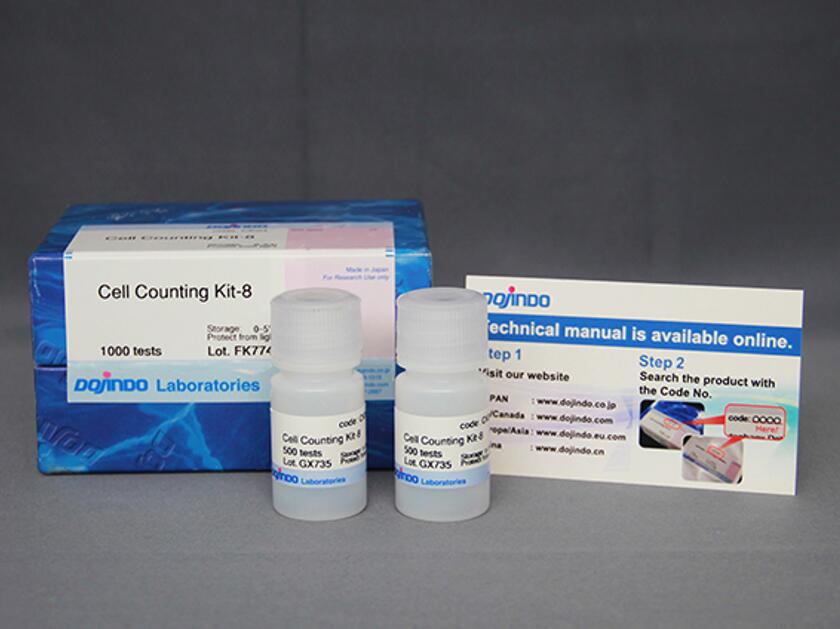
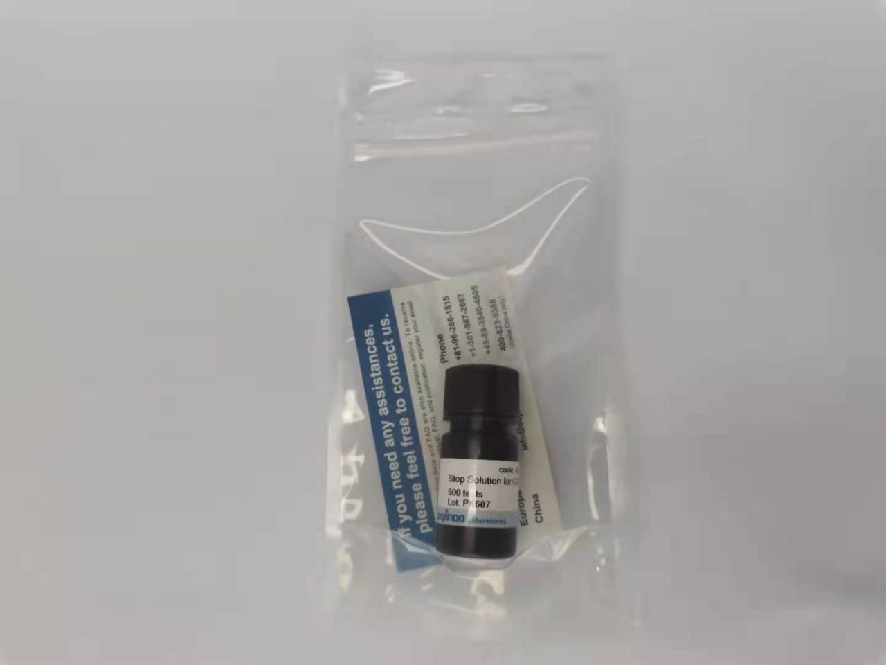
试剂盒内含

1622167990553553.jpg

产品概述

    1606975956235851.jpg

细胞毒性检测通常通过检测活细胞数量或者死细胞数量来确定。而为了更加全面的评价细胞毒性情况,多种评价方法相互验证的需求与日俱增。Viability/CytotoxicityMultiplex Assay Kit可以综合评价细胞毒性。试剂盒内含Cell Counting Kit-8 (CCK-8) 和Cytotoxicity LDH Assay Kit-WST® (LDH Assay Kit),分别用于测定活细胞和死细胞数量。

CCK-8通过检测活细胞中脱氢酶活性来确定活细胞数,而LDH Assay Kit通过检测受损细胞膜释放LDH的量来确定死细胞数。LDH Assay Kit可以直接添加到含有细胞的培养基中检测 (一步法)。也可以分离细胞后,加到细胞培养基上清液中检测 (低损伤法,分离的细胞可以做其它实验)。两种细胞毒性评价指标,CCK-8和LDH Assay Kit(低损伤法) 可以使用同一批细胞同时进行实验。

产品特点

不同细胞毒性指标,双重印证。增加结果可信性。

单独检测CCK-8,细胞毒性判断不准确 2种指标一起检测,增加数据准确性
1、单纯检测活细胞数量(CCK-8、MTT、MTS、XTT等) 1、单纯检测活细胞数量(CCK-8、MTT、MTS、XTT等)
2、活细胞检测+死细胞检测(细胞膜损伤)

1622168130818113.jpg

检测原理

LDH Assay Kit:细胞膜损伤,LDH外漏,通过检测LDH活性判断细胞毒性。

CCK-8:可以使用NADH作为指标来确认活细胞中的代谢活性。

1606976532212969.jpg

同时检测的操作方法

1606976594787617.jpg

本试剂盒包括用于测量活细胞的CCK-8和用于测量死细胞的LDH分析试剂盒,两者均可通过平板分析法在相同的吸收波长下进行评估。 此外,同一细胞样本可用于同时评估活细胞和死细胞,从而节省了时间和精力。

性价比高

本产品作为CCK-8(500 tests)与Cytotoxicity LDH Assay Kit(500 tests)组合套装,低于单独购买的总价。适合新接触细胞毒性检测的用户,用于前期摸索实验条件。单独产品链接见下方。大包装购买,单价会降低很多。

Cell Counting Kit-8

Cytotoxicity LDH Assay Kit-WST

参考文献

1. Integrated Omics Reveals Tollip as an Regulator and Therapeutic Target for Hepatic Ischemia-Reperfusion Injury in Mice,

Hepatology., 2019, doi: 10.1002/hep.30705

2. Amplified Cancer Immunotherapy of Surface Engineering Antigenic Microparticles Vaccine by Synergistically Modulating Tumor

Microenvironment, ACS Nano, 2019, 13, 11, 12553-12566

3. A novel IFNα-induced long noncoding RNA negatively regulates immunosuppression by interrupting H3K27 acetylation in head

and neck squamous cell carcinoma, Molecular Cancer, 2020,19, 4, doi: 10.1186/s12943-019-1123-y

4. Untangling the co-effects of oriented nanotopography and sustained anticoagulation in a biomimetic intima on neovessel

remodeling, Biomaterials, 2019, 119654

5. In Vitro and in Vivo Analysis of Mineralized Collagen-Based Sponges Prepared by a Plasma- and Precursor-Assisted

Biomimetic Process, ACS Applied Materials & Interfaces, 2017, 9, 27, 22185-22194

6. MoS2-LA-PEI nanocomposite carrier for real-time imaging of ATP metabolism in glioma stem cells co-cultured with

endothelial cells on a microfluidic system, Biosensors and Bioelectronics, 2018, 99, 142-149

7. Biocompatible carbon nanotube fiber for implantable supercapacitors, Carbon, 2017, 122, 162-167

8. Genomic sequencing and editing revealed the GRM8 signaling pathway as potential therapeutic targets of squamous cell

lung cancer, Cancer Letters, 2019, 442, 53-67

9. Knockdown of TGF-β1 expression in human umbilical cord mesenchymal stem cells reverts their exosome-mediated EMT

promoting effect on lung cancer cells, Cancer Letters, 2018, 428:34-44

10. Restriction of exogenous DNA expression by SAMHD1, Science Bulletin, 2020, https://doi.org/10.1016/j.scib.2019.12.028

11. PARP1 inhibition alleviates injury in ARH3-deficient mice and human cells, JCI Insight, 2019, 4(4), e124519

12. Downregulation of MCL-1 and upregulation of PUMA using mTOR inhibitors enhance antitumor efficacy of BH3 mimetics in

triple-negative breast cancer, Cell Death & Disease, 2018, 9(2), 137

13. Ischemic preconditioning attenuates ischemia/reperfusion-induced kidney injury by activating autophagy via the SGK1

signaling pathway, Cell Death & Disease, 2018, 9, 338

14. Pathological Endogenous a-Synuclein Accumulation in Oligodendrocyte Precursor Cells Potentially Induces Inclusions in

Multiple System Atrophy, Stem Cell Reports, 2018, 10(2), 356-365

15. PDH-mediated metabolic flow is critical for skeletal muscle stem cell differentiation and myotube formation during regeneration

in mice, FASEB Journal, 2019, 33(7), 8094-8109

16. Benzo[a]pyrene-decreased gap junctional intercellular communication via calcium/calmodulin signaling increases apoptosis in

TM4 cells, Journal of Applied Toxicology, 2018, 38(8), 1091-1103

关联产品

Cell Counting Kit-8细胞增殖毒性检测试剂盒CCK-8(CCK8)
细胞增殖-毒性检测试剂盒CCK-8(CCK8)
LDH检测试剂盒—Cytotoxicity LDH Assay Kit-WST
乳酸脱氢酶(LDH)检测试剂盒
Cell Counting Kit-Luminescence试剂盒
细胞增殖/毒性检测试剂盒-发光法(CCK-L)
Cell Count Normalization Kit试剂盒
细胞计数标准化试剂盒
Stop Solution for CCK-8试剂
CCK-8反应终止液
在线小工具

实验工具|稀释计算器|摩尔浓度计算器

Hampton蛋白结晶试剂盒LCP Lipidic Cubic Phase Screening

上海金畔生物代理Hampton research品牌蛋白结晶试剂耗材工具等,我们将竭诚为您服务,欢迎访问Hampton research官网或者咨询我们获取更多相关Hampton research品牌产品信息。Products > 96 Well Crystallization Plates > Swissci > LCP Lipidic Cubic Phase Screening

LCP Lipidic Cubic Phase Screening

Applications

  • Lipidic cubic phase crystallization

Features

  • UVP cover film (UV compatible)
  • No cross contamination
  • Membrane protein crystallization
  • 6 mm diameter drop well area
  • 0.1 mm (100 μm) deep drop well when LCP plate is sealed
  • Maximum drop volume is 1.6 microliters per well

Description

The Lipidic Cubic Phase Crystallization (LCP) plate facilitates the automation and increased throughput of LCP crystallization set-ups. This novel system enables LCP screening to be performed accurately and with ease – using manual – or automated systems to complete the delivery of the solutions.

Working with innovative scientists Professor Gebhard Schertler and Pat Edwards from the MRC laboratory in Cambridge, Swissci AG developed a new system for setting up LCP screens.

The Swissci LCP plate ensures crystallography set-up is fast, cost effective and robotically adaptable. In addition Ultra Violet light can be used with the glass / UVP polymers to visualize proteins without the light scattering associated with traditional polymeric solutions.

The LCP technique for crystallizing membrane proteins can be difficult and time-consuming to set up as it utilizes highly viscous lipid mesophases to reconstitute proteins. Swissci AG and MRC overcame these problems with the LCP plate. This product combines an easy to use sandwich system with dedicated coupling tapes and thin film UVP cover plates. It conforms to SBS standards and thus is perfect for modern automation applications where high throughput is required.

The Advantages of the LCP plate
• Ready to use the plate fits laboratory robotics and uses a Swissci unique polymer to ensure UV visualization is not compromised by polarization.
• Easy sealing with dry tabbed adhesive tape exposure and thin UVP cover film. Included in the kit is a plate leveling device to ensure the cover is perfectly adhered to the bottom LCP plate.
• No cross contamination and SBS standard.
• Unique low tack plate security allows for the sandwich plate to be removed from the base plate when required – in-situ x-ray data collection and structure determination is then enabled.

Here’s a description of the LCP plate from bottom to top. a) An SBS standard base plate frame support. b) 0.7 mm thick UVP plastic slide drop support containing all 96 drop wells with 0.1 mm thick layer of low tack sealant (no sealant in drop area). Flat, circular, 6 mm diameter drop wells to contain the LCP, sample and reagent. c) Removable brown/tan paper protects sealant until plate is ready to use. d) 0.1 mm thick UVP sealing film. e) Removable protective dust cover. The kit is completed with a Swissci plate leveling device.

To use the LCP plate, pipette LCP, sample and reagent into the 96 drop wells. Peel back and remove the brown/tan paper to expose the adhesive. Place the sealing film onto the LCP plate. Remove the protective film for viewing.

Maximum drop volume is 1.6 microliters per well.

Show More


LCP Lipidic Cubic Phase Screening

Click to Zoom In

LCP Lipidic Cubic Phase Screening

CAT NO

HR3-186

NAME

Lipidic Cubic Phase Screening Kit

DESCRIPTION

20 plate case

PRICE

$422.00

Temporarily Unavailable
More Available Soon!

Support Material(s)

LCP Lipidic Cubic Phase Screening HR3-186 LCP Lipidic Cubic Phase Screen Kit User Guide

Hampton蛋白结晶试剂盒Diaplate

上海金畔生物代理Hampton research品牌蛋白结晶试剂耗材工具等,我们将竭诚为您服务,欢迎访问Hampton research官网或者咨询我们获取更多相关Hampton research品牌产品信息。Products > 96 Well Crystallization Plates > Swissci > Diaplate

Diaplate

Applications

  • Protein and peptide purification/concentration
  • Variation of crystallisation conditions for crystal growth
  • Removal of dyes after protein labelling
  • Electro-elution of proteins
  • Protein and DNA desalting/buffer exchange
  • Protein in vitro translation
  • Glycoprotein modification and engineering
  • Enzyme activity/binding assays/detergent removal

Features

  • Protein crystallisation applications allow for unique matrix changing
  • Simple handling, ready to use, easy sample recovery
  • Complete kit, no additional materials required
  • Complies with ANSI/SBS standards for automation
  • Pipetting can be performed manually or using a robot
  • Ideal for the desalting of up to 3.2µl protein solution
  • No cross contamination or leakage between wells guaranteed
  • 96 individual regenerated cellulose membranes; sulphur and heavy metal free
  • Molecular weight cut off of 10,000 Daltons
  • Low protein binding and high protein retention
  • Gentle and efficient desalting without disturbing valuable protein complexes
  • High throughput; simultaneous processing of 96 experiments
  • Reliable and uniform, stable construction
  • Single use – disposable device; no cleaning required

Description

The disposable Diaplate is a 96 well micro-dialysis plate ideal for the desalting of protein from very small volumes of up to a maximum of 3.2 µl.

The sample solution is pipetted into each of the 96 wells with up to 0.35ml dialysis solution used for exchange. The Diaplate is ANSI/SLAS 1 2004 standards providing high throughput use with robotic dispensing. Dialysis is recommended for 2-3 hours and then the dialysis buffer is changed being left for another 2-3 hours to achieve over 90% exchange. Further dialysis steps can be carried out as required. Incubation, rocking table for speed and moisture chamber for full humidification can be applied to the experiment if desired.

Each of the 96 wells has a separate regenerated cellulose membrane inside meaning no cross contamination or leakage between wells. The size of the compounds that pass through the membrane are determined by its Molecular Weight Cut Off (MWCO). The Swissci Diaplate comes as standard with a specially selected membrane that has a MWCOs of 10,000 Daltons. The Diaplate kit includes: Diaplate with 200 Micron Pressure Adhesive Spacer, 200 Micron UVP Cover Film, engraved UV Protective Cover and a sealing Paddle. Swissci W82010.

Diaplate Specifications
Membrane molecular weight cut-off (MWCO) 10kDa (10,000 Daltons)
Maximum sample capacity 3.2 µl
Maximum dialysate or reagent capacity 350 µl
Plate dimensions 127.76 +/- 0.5 x 85.48 +/- 0.5 mm

Show More


Diaplate

Click to Zoom In

Diaplate
Diaplate
Diaplate
Diaplate

CAT NO

HR3-253

NAME

Diaplate

DESCRIPTION

10 kDa MWCO 3/pack

PRICE

$136.00

cart quote

Support Material(s)

Diaplate HR3-253 Diaplate User GuideDiaplate HR3-253 Diaplate Product Information

Cell Counting Kit-Luminescence试剂盒 CK18 ATP活性检测 200 tests


品名 货号 用途 规格 价格
Glucose(葡萄糖)摄取能力检测试剂盒-Blue UP01 葡萄糖摄取能力检测(蓝色荧光) 1 set 3980.00
Glucose(葡萄糖)摄取能力检测试剂盒-Green UP02 葡萄糖摄取能力检测(绿色荧光) 1 set 3980.00
Glucose(葡萄糖)摄取能力检测试剂盒-Red UP03 葡萄糖摄取能力检测(红色荧光) 1 set 3980.00
氨基酸摄取能力检测试剂盒——Amino Acid Uptake Assay Kit UP04 检测细胞摄取氨基酸的能力 100tests 2980.00
胱氨酸摄取能力检测试剂盒—Cystine Uptake Assay Kit UP05 胱氨酸摄取能力检测 100tests 3980.00

订购以下任意代谢类产品,即可获得100元礼品~

(点击产品,即可进入产品详情链接)

品名 货号 用途 规格 价格
Cell Counting Kit-Luminescence试剂盒 CK18 ATP活性检测 200 tests 620.00
Lipi-Blue试剂 LD01 脂滴检测(蓝色) 10 nmol 1940.00
Lipi-Green试剂 LD02 脂滴检测(绿色) 10 nmol 1940.00
Lipi-Red试剂 LD03 脂滴检测(红色) 10 nmol 1940.00
Lipi-Deep Red试剂 LD04 脂滴检测(深红色) 10 nmol 1940.00
Lipid Droplet Assay Kit-Blue试剂 LD05 脂滴荧光检测(蓝色) 1set 2980.00
Lipid Droplet Assay Kit-Deep Red试剂 LD06 脂滴荧光检测(深红色) 1set 2980.00
Lactate Assay Kit-WST试剂盒 L256 乳酸检测试剂盒 50 tests 1980.00
200 tests 6770.00
NAD/NADH Assay Kit-WST试剂盒 N509 NAD/NADH检测试剂盒 100 tests 5840.00
NADP/NADPH Assay Kit-WST试剂盒 N510 NADP/NADPH检测试剂盒 100 tests 5840.00
Glucose Assay Kit-WST试剂盒 G264 葡萄糖含量检测 50 tests 1940.00
200 tests 4090.00
Glutamine Assay Kit-WST试剂盒 G268 谷氨酰胺的定量检测 1set 6060
Glutamine Assay Kit-WST试剂盒 G269 谷氨酸的定量检测 1set 5500.00
糖酵解/氧化磷酸化检测试剂盒—Glycolysis/OXPHOS Assay Kit G270 方便快捷的检测糖酵解能、细胞代谢途径转移、细胞对糖酵解途径和氧化磷酸化途径的依赖程度 50 tests 5100.00
ADP/ATP比率检测试剂盒—ADP/ATP Ratio Assay Kit-Luminescence A552 检测细胞中ADP与ATP的比率 100 tests 4100.00
氨基酸摄取能力检测试剂盒——Amino Acid Uptake Assay Kit UP04 检测细胞摄取氨基酸的能力 20tests 1130.00
胱氨酸摄取能力检测试剂盒—Cystine Uptake Assay Kit UP05 胱氨酸摄取能力检测 20tests 1490.00

心动不如行动、赶快来订购吧~

Hampton蛋白结晶试剂盒Vial Clamp™ – Curved/HR4-671/HR4-672

上海金畔生物代理Hampton research品牌蛋白结晶试剂耗材工具等,我们将竭诚为您服务,欢迎访问Hampton research官网或者咨询我们获取更多相关Hampton research品牌产品信息。

Products > Cryocrystallography > Cryo Tools > Vial Clamp™ – Curved

Vial Clamp™ – Curved

应用

  • Vial support & manipulation
  • 样品瓶支持和操作

特征

  • Curved end tip
  • Hemostat style closure
  • 弯曲端头
    止血式封口

描述

The Vial Clamp – Curved is a chrome plated, hemostat style tool. It has a tip shaped to hold the storage vial of all the CrystalCap Systems at an angle when the clamp is closed. The clamp can be locked using the hemostat style closure. The end of the clamp, where the vial is held, is curved at either a 45°/135° or 110°/ 70° angle. When the vial is placed in the clamp, the length of it is positioned to the clamp at either of those same angles. The overall length of each clamp is the same, 195 mm.

Vial Clamp – Curved 是一种镀铬的止血工具。 它有一个尖端,可在夹子闭合时以一定角度固定所有 CrystalCap 系统的存储瓶。 可以使用止血器式闭合来锁定夹子。 夹持小瓶的夹具末端以 45°/135° 或 110°/70° 角弯曲。 当小瓶放置在夹具中时,它的长度以这些相同角度中的任何一个定位到夹具。 每个夹具的总长度相同,均为 195 毫米。

HR4-671 Vial Clamp Curved 45°/135°

Vial Clamp™ - Curved
CAT NO

HR4-671

NAME

Vial Clamp – Curved 45°/135°

DESCRIPTION

each

PRICE

$67.00

CAT NO

HR4-672

NAME

Vial Clamp – Curved 110°/70°

DESCRIPTION

each

PRICE

$97.00

相关项目

  • CrystalCap™
  • CrystalCap™ SPINE HT

线粒体

线粒体

线粒体(mitochondrion) 是一种存在于大多数细胞中的由两层膜包被的细胞器,是细胞中制造能量的结构,是细胞进行有氧呼吸的主要场所,线粒体拥有自身的遗传物质和遗传体系,但其基因组大小有限,是一种半自主细胞器。除了为细胞供能外,线粒体还参与诸如细胞分化、细胞信息传递和细胞凋亡等过程,并拥有调控细胞生长和细胞周期的能力。最近越老越多的研究发现线粒体在细胞中的作用远远不止”细胞能量站”。它们参与了各种细胞功能调控,与很多人类疾病存在着莫大的联系。包括细胞信号传导、代谢、自噬、衰老和肿瘤发生都与线粒体的质量和活性相关
线粒体损伤
线粒体自噬
线粒体氧化应激
线粒体染色
品名 货号 用途
线粒体膜电位检测试剂盒 MT13 线粒体膜电位检测
线粒体膜电位检测试剂盒—JC-1 MitoMP Detection Kit MT09 线粒体膜电位检测
Cellstain- MitoRed试剂 R237 线粒体ATP检测-红色
Cellstain- Rh123试剂(罗丹明123) R233 线粒体ATP检测-绿色
品名 货号 用途
Mtphagy Dye试剂 MT02 线粒体自噬
线粒体自噬—Mitophagy Detection Kit MD01 线粒体自噬检测
品名 货号 用途
mtSOX Deep Red – Mitochondrial Superoxide Detection MT14 线粒体超氧化物检测
铁离子荧光探针—Mito-FerroGreen M489 线粒体内二价铁离子检测
Si-DMA for Mitochondrial Singlet Oxygen Imaging试剂 MT05 线粒体内单线态氧检测
MitoPeDPP试剂 M466 线粒体内脂质过氧化物检测
品名 货号 用途
MitoBright IM Red for Immunostaining试剂 MT15 免疫荧光用线粒体荧光染料Red
MitoBright LT Green试剂 MT10 线粒体长效荧光染色(绿色)
MitoBright LT Red试剂 MT11 线粒体长效荧光染色(红色)
MitoBright LT Deep Red试剂 MT12 线粒体长效荧光染色(深红色)

线粒体功能研究

image.png

线粒体简要通路图海报下载

同仁化学 线粒体简要通路图.pdf

线粒体相关检测试剂

线粒体自噬检测

线粒体自噬
试剂 Mtphagy Dye Keima-Red
原理 线粒体自噬染料是一种PH敏感的荧光探针,该染料聚集在线粒体中,并由溶酶体的酸性条件而发出荧光 这是一种基于PH感应比值的荧光蛋白。该蛋白在溶酶体中具有比较高的荧光比值(如550 nm/440 nm)。
固定细胞染色
活细胞染色 Yes Yes
活细胞染色后固定
染色时间 >30 min
Ex/Em 530/700 440,550/620
产品货号 MD01 , MT02

线粒体膜电位检测

Membrane potential

线粒体膜电位

试剂 JC-1 TMRM,   TMRE
原理 JC-1是一种被广泛使用的小分子线粒体膜电位探针,依赖于线粒体膜电位在线粒体中聚集,染料伴随聚集过程,荧光从绿色   (530 nm) 变为红色 (590 nm)。当线粒体发生去极化,红/绿荧光强度比值降低。 该试剂是细胞渗透性荧光染料,由于膜电位在完整的线粒体中积累。探针扩散发生在膜电位降低的受损线粒体中。
固定细胞染色
活细胞染色 Yes Yes
活细胞染色后固定
染色时间 10- 60 min 30- 60 min
Ex/Em Monomer:514/529 550/575
J-aggregation: 585/590
产品货号 MT09

线粒体金属离子检测

Iron ion (Fe2+)

亚铁离子

Calcium ion (Ca2+)

钙离子

试剂 Mito-FerroGreen Rhod 2-AM
原理 该试剂是一种细胞通透性探针,其积累在线粒体中,并与线粒体中的亚铁离子发生特异性反应,发出绿色荧光。 该试剂是一种细胞通透性探针,该探针积聚在线粒体中,并与线粒体中的钙离子发生特异性反应,发出红色荧光。
固定细胞染色
活细胞染色 Yes Yes
活细胞染色后固定
染色时间 30 min 30-60 min
Ex/Em 505/535 553/576
产品货号 M489 R002

线粒体荧光染色

Mitochondria staining

线粒体染色

试剂 MitoBright LT series MitoTracker series
原理 细胞渗透性荧光染料,基于线粒体膜电位而在完整的线粒体中积累。 细胞渗透性荧光染料,基于线粒体膜电位而在完整的线粒体中积累。
固定细胞染色
活细胞染色 Yes Yes
活细胞染色后固定
染色时间 >10 min 15 -45 min
Ex/Em 493/508,547/563, 643/663 490/516~644/665
产品货号 MT10、MT11、MT12
Mitochondria staining

线粒体染色

试剂 DsRed MitoRed Rh123
原理 一种红色荧光蛋白染料,通过转染试剂与完整的线粒体结合。 细胞渗透性荧光染料,基于线粒体膜电位而在完整的线粒体中积累。 细胞渗透性荧光染料,基于线粒体膜电位而在完整的线粒体中积累。
固定细胞染色
活细胞染色 Yes Yes Yes
活细胞染色后固定
染色时间 Expression in 8 -12 hrs. >15 min >15 min
Ex/Em 558/583 560/580 507/529
产品货号 R237 R233

线粒体氧化应激

Lipophilic peroxide

脂质过氧化物

Singlet oxygen

单线态氧

Superoxide

超氧化物

试剂 MitoPeDPP Si-DMA MitoSOX
原理 MitoPeDPP是一种新型荧光染料,由于其具有三苯基膦结构,因此可以穿过细胞膜并在线粒体中聚集。聚集在线粒体内膜上的MitoPeDPP可以被脂质过氧化物氧化而释放出强荧光。 该试剂是一种细胞渗透性荧光探针,积聚在线粒体中,与线粒体中产生的单线态氧发生反应,发出红色荧光。 该试剂是一种细胞渗透荧光探针,积聚在线粒体中,与线粒体中产生的超氧化物反应,发出红色荧光。
固定细胞染色
活细胞染色 Yes Yes Yes
活细胞染色后固定
染色时间 > 15 min > 45 min > 10 min
Ex/Em 452/ 470 644/ 670 510/ 590
产品货号 M466 MT05

引用论文

① H. Tanaka, S. Takebayashi, A. Sakamoto, N. Saitoh, S. Hino and M. Nakao, “The SETD8/PR-Set7 Methyltransferase Functions as a Barrier to Prevent Senescence-Associated Metabolic Remodeling.”, Cell Reports, 2017, 18(9), 2148.
② L. Garcia-Prat, M. Martinez-Vicente and P. Munoz-Canoves, “Autophagy: a decisive process for stemness”, Oncotarget, 2016, 7(11), 12286.
③ M. Bitar, S. Abdel-Halim and F. Al-Mulla, “Caveolin-1/PTRF upregulation constitutes a mechanism for mediating p53-induced cellular senescence: implications for evidence-based therapy of delayed wound healing in diabetes”, Am J Physiol Endocrinol Metab., 2013, 305(8), E951.
④ C. Wiley, M. Velarde, P. Lecot, A. Gerencser, E. Verdin, J. Campisi, et. al., “Mitochondrial Dysfunction Induces Senescence with a Distinct Secretory Phenotype”, Cell Metab., 2016, 23(2), 303.
⑤ E. Liao, Y. Hsu, Q. Chuah, Y. Lee, J. Hu, T. Huang, P-M Yang & S-J Chiu, “Radiation induces senescence and a bystander effect through metabolic alterations.”, Cell Death Dis., 2014, 5, e1255.
⑥ K. Nishimura, T. Kumazawa, T. Kuroda, A. Murayama, J. Yanagisawa and K. Kimura, “Perturbation of Ribosome Biogenesis Drives Cells into Senescence through 5S RNP-Mediated p53 Activation”, Cell Rep. 2015, 10(8), 1310.
⑦ M. J. Son, Y. Kwon, T. Son and Y. S. Cho, “Restoration of Mitochondrial NAD+ Levels Delays Stem Cell Senescence and Facilitates Reprogramming of Aged Somatic Cells”, Stem Cells. 2016, 34(12), 2840.

Cell Cycle Assay Solution Deep Red试剂货号:C548 细胞周期检测试剂盒-深红色

Cell Cycle Assay Solution Deep Red试剂货号:C548
细胞周期检测试剂盒-深红色
Cell Cycle Assay Solution Deep Red
商品信息
储存条件:0-5 ℃
运输条件:常温

特点:

·检测时间短

·固定和不固定都可以

·悬浮细胞和贴壁细胞都可以

下载说明书
SDS下载
选择规格:
50 tests
价格:
期货 
 
细胞周期
产品概述
产品特性
实验案例
常见问题Q&A
参考文献

产品概述

  细胞周期的调控系统与细胞增殖密切相关。一个细胞产生两个子细胞的细胞分裂是细胞周期的一部分。细胞周期可以通过流式细胞术测量细胞DNA染色,然后分析染色细胞核在细胞周期阶段的比例来分析。细胞周期分为两个主要阶段:间期和有丝分裂(M)期。间期由G1期(发生在细胞分裂之前的DNA合成准备)、S期(DNA合成和复制)和G2期(具有重复染色体的单个细胞)组成。M期包括有丝分裂和胞质分裂。细胞周期的进展受细胞周期蛋白和细胞周期蛋白依赖性激酶的调控。分析细胞周期检查点的作用机制,对确定抗癌药物及其他药物的作用具有重要意义

1607485076273864.png

产品特性

产品特点:检测时间短

碘化丙啶(PI)通常用于使用流式细胞仪进行细胞周期分析。与这种分析方法相比,深红色细胞周期测定溶液具有良好的膜渗透性,并使用对DNA具有高特异性的染料,因此仅需将试剂添加到细胞悬液中就可以分析细胞周期。此外,深红色细胞周期测定解决方案中还提供了633 nm激光器,使您可以同时使用高度通用的488 nm激光器。

1607485106948667.png

实验案例

对比实验:准确的判段活细胞的细胞周期

用细胞周期流式检测试剂(蓝色和深红色)染色的活CHO细胞,同时使用广泛应用的PI染色产品和同仁已有的细胞周期检测试剂盒,进行了类似的实验。结果,通过细胞周期流式检测试剂获得的结果与PI染色结果相同(如下所示)。与四种不同的产品相比,我们的产品在活细胞中获得了清晰的直方图峰。

1607484701403788.png

示例:抗癌剂引起的细胞功能的变化

阿霉素(DOX)在细胞周期的G2 / M期抑制细胞增殖,并诱导细胞衰老。将DOX加入A549细胞后,G2 / M期的峰值增高(Cell Cycle Assay Solution Deep Red、Blue;C548\C549),诱导细胞衰老(Cellular Senescence Detection Kit – SPiDER-βGal;SG03)和线粒体膜电位发生变化(JC-1 MitoMP Detection Kit;MT09)

1607484736305104.png

常见问题Q&A

Q1:悬浮细胞和贴壁细胞都可以检测吗?
A1:悬浮细胞和贴壁细胞都可以使用。
Q2:胰酶会影响检测吗?
A2:如果实验中有胰酶残留,会影响检测;

如果回收细胞时使用胰酶,请确保按照方案进行清洁操作,去除残留胰酶。

Q3:需要固定细胞吗?
A3:固定和不固定细胞都可以。
Q4:如果需要对要检测的固定细胞进行保存,是应该染色前保存还是染色后保存?
A4:都可以,本公司实际对比案例:

分别在保存固定细胞前后做了染色实验,检测结果没有差异

・将固定细胞冷藏(4℃)后保存1周,根据使用说明书对细胞进行染色。

・根据使用说明书对固定细胞进行染色,将细胞冷藏(4℃)后保存1周。

*冷冻保存细胞的话有可能会沉淀不溶物,所以不推荐冷冻保存。

Q5:用Cell Cycle Assiay  Soluton染色后,为了除去过量的试剂可以清洗细胞吗?
A5:不推荐染色后清洗细胞。

由于洗涤时的离心操作,有可能影响细胞周期解析的实验结果。

Q6:做流式检测时,应该注意哪些方面呢?
A6:流式检测时,需要在合适的条件下进行测检测。

以下列举了一些失败的案例。

1、检测通道选择不合适

例如一般的细胞周期检测都会使用PI(PE)通道,但是我们试剂盒选择PI通道并不能得到最适结果,请参照一下波段选择最适通道:

2、检测时电压过高(或过低)。

当流式检测时电压过高(或过低)时,都无法得到最适结果。

请通过直方图将G0/G1期、S期、G2/M期调整为清晰的测量电压。

3、细胞数量选择不合适

例如流式检测是圈门选择不恰当,会导致结果不好。

分析时请对选择所有细胞。

参考文献

1)細胞(HCAECs)

N. Sasaki, Y. Itakura and M. Toyoda, “Rapamycin promotes endothelial-mesenchymal transition during stress-induced premature senescence through the activation of autophagy”, Cell Commun. Signal, 2020, 18(1), 43

2)細胞(ARPE-19)

T. Yamazaki, H. Suzuki, S. Yamada, K. Ohshio, M. Sugamata, T. Yamada and Y. Morita, “Lactobacillus paracasei KW3110 Suppresses Inflammatory Stress-Induced Premature Cellular Senescence of Human Retinal Pigment Epithelium Cells and Reduces Ocular Disorders in Healthy Humans”, Int J Mol Sci, 2020, 21(14), 5091

2)細胞(MIA PaCa-2)

N. Sasaki, F. Gomi, F. Hasegawa, K. Hirano, M. Fujiwara, M. Toyoda and T. Ishiwata, “Characterization of the metastatic potential of the floating cell component of MIA PaCa-2, a human pancreatic cancer cell line”, Biochem. biophys. res. commun, 2020, 522(4), 881-888

关联产品

细胞周期检测试剂盒-PI/RNase Staining
细胞周期检测试剂盒
Cell Cycle Assay Solution Blue试剂
细胞周期检测试剂盒-蓝色
在线小工具

实验工具|稀释计算器|摩尔浓度计算器

Cell Cycle Assay Solution Blue试剂货号:C549 细胞周期检测试剂盒-蓝色

Cell Cycle Assay Solution Blue试剂货号:C549
细胞周期检测试剂盒-蓝色
Cell Cycle Assay Solution Blue
商品信息
储存条件:0-5 ℃
运输条件:常温

特点:

·检测时间短

·固定和不固定都可以用

·悬浮细胞核贴壁细胞都可以用

下载说明书
SDS下载
选择规格:
50 tests
期货 
 
细胞周期
产品概述
产品特性
实验案例
常见问题Q&A

产品概述

细胞周期的调控系统与细胞增殖密切相关。一个细胞产生两个子细胞的细胞分裂是细胞周期的一部分。细胞周期可以通过流式细胞术测量细胞DNA染色,然后分析染色细胞核在细胞周期阶段的比例来分析。细胞周期分为两个主要阶段:间期和有丝分裂(M)期。间期由G1期(发生在细胞分裂之前的DNA合成准备)、S期(DNA合成和复制)和G2期(具有重复染色体的单个细胞)组成。M期包括有丝分裂和胞质分裂。细胞周期的进展受细胞周期蛋白和细胞周期蛋白依赖性激酶的调控。分析细胞周期检查点的作用机制,对确定抗癌药物及其他药物的作用具有重要意义

blob.png

产品特性

产品特点:检测时间短

碘化丙啶(PI)通常用于使用流式细胞仪进行细胞周期分析。与这种分析方法相比,深红色细胞周期测定溶液具有良好的膜渗透性,并使用对DNA具有高特异性的染料,因此仅需将试剂添加到细胞悬液中就可以分析细胞周期。此外,深红色细胞周期测定解决方案中还提供了633 nm激光器,使您可以同时使用高度通用的488 nm激光器。

1607485027827020.png

实验案例

对比实验:准确的判段活细胞的细胞周期

用细胞周期流式检测试剂(蓝色和深红色)染色的活CHO细胞,同时使用广泛应用的PI染色产品和同仁已有的细胞周期检测试剂盒,进行了类似的实验。结果,通过细胞周期流式检测试剂获得的结果与PI染色结果相同(如下所示)。与四种不同的产品相比,我们的产品在活细胞中获得了清晰的直方图峰。

1607484701403788.png

示例:抗癌剂引起的细胞功能的变化

阿霉素(DOX)在细胞周期的G2 / M期抑制细胞增殖,并诱导细胞衰老。将DOX加入A549细胞后,G2 / M期的峰值增高(Cell Cycle Assay Solution Deep Red、Blue;C548\C549),诱导细胞衰老(Cellular Senescence Detection Kit – SPiDER-βGal;SG03)和线粒体膜电位发生变化(JC-1 MitoMP Detection Kit;MT09)

1607484736305104.png

常见问题Q&A

Q1:悬浮细胞和贴壁细胞都可以检测吗?
A1:悬浮细胞和贴壁细胞都可以使用。
Q2:胰酶会影响检测吗?
A2:如果实验中有胰酶残留,会影响检测;

如果回收细胞时使用胰酶,请确保按照方案进行清洁操作,去除残留胰酶。

Q3:需要固定细胞吗?
A3:固定和不固定细胞都可以。
Q4:如果需要对要检测的固定细胞进行保存,是应该染色前保存还是染色后保存?
A4:都可以,本公司实际对比案例:

分别在保存固定细胞前后做了染色实验,检测结果没有差异

・将固定细胞冷藏(4℃)后保存1周,根据使用说明书对细胞进行染色。

・根据使用说明书对固定细胞进行染色,将细胞冷藏(4℃)后保存1周。

*冷冻保存细胞的话有可能会沉淀不溶物,所以不推荐冷冻保存。

Q5:用Cell Cycle Assiay  Soluton染色后,为了除去过量的试剂可以清洗细胞吗?
A5:不推荐染色后清洗细胞。

由于洗涤时的离心操作,有可能影响细胞周期解析的实验结果。

Q6:做流式检测时,应该注意哪些方面呢?
A6:流式检测时,需要在合适的条件下进行测检测。

以下列举了一些失败的案例。

1、检测通道选择不合适

例如一般的细胞周期检测都会使用PI(PE)通道,但是我们试剂盒选择PI通道并不能得到最适结果,请参照一下波段选择最适通道:

2、检测时电压过高(或过低)。

当流式检测时电压过高(或过低)时,都无法得到最适结果。

请通过直方图将G0/G1期、S期、G2/M期调整为清晰的测量电压。

3、细胞数量选择不合适

例如流式检测是圈门选择不恰当,会导致结果不好。

分析时请对选择所有细胞。

关联产品

细胞周期检测试剂盒-PI/RNase Staining
细胞周期检测试剂盒
Cell Cycle Assay Solution Deep Red试剂
细胞周期检测试剂盒-深红色

细胞周期检测试剂盒-PI/RNase Staining货号:C543 Cell Cycle Assay Kit – PI/RNase Staining

细胞周期检测试剂盒-PI/RNase Staining货号:C543
细胞周期检测试剂盒
Cell Cycle Assay Kit – PI/RNase Staining
商品信息
储存条件:0-5度保存,避光
运输条件:室温

特点:

·日本原装进口试剂盒

·固定和不固定都可使用

下载说明书
SDS下载
选择规格:
50tests
现货 
 
细胞周期
活动进行中
试剂盒内含
产品概述
所需的设备和材料
溶液制备
基本操作
常见问题Q&A

活动进行中

订购满5000元,200元礼品等你拿

凑单关联产品TOP5

NO.1.    Annexin V, FITC Apoptosis Detection Kit    细胞凋亡检测

NO.2.    Cell Counting Kit-8    细胞增殖毒性检测

NO.3.    ROS Assay Kit -Highly Sensitive DCFH-DA-    ROS检测

NO.4.    Cellular Senescence Detection Kit    细胞衰老检测

NO.5.    Annexin V, 633 Apoptosis Detection Kit    细胞凋亡检测

试剂盒内含

1622173397780997.jpg

产品概述

  DNA含量分析在流式细胞检测中,占有相当大的比例。通过这种方法,研究者可以进行抗癌药物的毒性评估,并对恶性肿瘤细胞的预后效果及进展程度进行分析。在细胞周期检测中,DNA含量也同样作为重要的分析手段而被广泛应用。通过细胞DNA含量的测定,可计算各细胞周期的百分率,也可通过DNA片段化检测、DNA双染标记等方法检测细胞凋亡。

Cell Cycle Assay Kit-PI/RNase Staining试剂盒,可快速完成细胞周期的分析,操作简便。本产品使用碘化丙啶 (PI) 作为DNA荧光染料,通过流式细胞术得到细胞周期中G0/G1,G2和S期细胞的比例。

所需的设备和材料

– 流式细胞仪(碘化丙啶PI:λex=535 nm,λem=617 nm)

– 37℃培养箱

– PBS缓冲液

– 1.5 ml微量管

– 100-1000 μl、20-200 μl、2-20 μl移液器

溶液制备

本产品在固定细胞及不细胞固定的情况下皆可使用。

配置Working solution (1 sample)

取500 μl Assay Buffer 后,分别加入25 μl PI Solution和2.5 μl RNase Solution。

*工作液易遇光分解,请在使用前立即配置并用铝箔纸包裹避光保存。

配置好的工作液1天内用完。

基本操作

非固定细胞

1. 将细胞密度调整为1-5×10⁶cells/ml a)

2. 取1 ml的细胞悬液加入到1.5 ml微型管中,在1,000×g离心3 min b)

3. 去除上清液后加入1 ml PBS缓冲液清洗细胞,在1,000×g离心3 min。

4. 去除上清液后加入0.5 ml Working Solution。

5. 在4℃避光培养30 min。

6. 涡旋振荡使细胞分散,在37℃避光培养30 min。

7. 涡旋振荡使细胞分散,用尼龙筛网过滤,以去除样品中的细胞团块 c)

8. 用流式细胞仪检测。

a) 贴壁细胞用胰蛋白酶-EDTA消化细胞后,用PBS洗涤并离心收集细胞。

b) 根据不同的细胞种类,需适当调整转速与离心时间。

c) 样品处理后,剩余部分无法继续保存并使用。

固定细胞

1. 将细胞密度调整为1-5×10⁶cells/ml a)

2. 取1 ml的细胞悬液加入到1.5 ml微型管中,在1,000×g 离心3 min。

3. 去除上清,并在细胞团中加入1 ml的-20℃保存的70%乙醇。

4. 涡旋振荡使细胞分散,在4℃下静置2 h b)

5. 在1,000×g 离心3 min后,去除乙醇 c)

6. 加入1 ml PBS缓冲液清洗细胞,在1,000×g 离心3 min。

7. 去除上清液后加入0.5 ml Working Solution。

8. 涡旋振荡使细胞分散,在37℃避光培养30 min。

9. 涡旋振荡使细胞分散,在4℃避光培养30 min。

10. 涡旋振荡使细胞分散,用尼龙筛网过滤,以去除样品中的细胞团块 d)

11. 用流式细胞仪检测。

a) 贴壁细胞用胰蛋白酶-EDTA消化细胞后,用PBS洗涤并离心收集细胞。

b) 根据不同的细胞种类,需适当调整固定时间。

c) 根据不同的细胞种类,需适当调整转速与离心时间。

d) 样品处理后可以在4℃以下保存并继续使用,但是保存时间长短与细胞种类有关。

常见问题Q&A

Q1、C543用于分散细胞团聚的筛网的品牌、型号信息?
A1:FALCON 5mL Polystyrene Round-Bottom Tube with Cell-Strainer Cap 型号:352235
Q2:实验需要在37度、4度环境中分别培养30min,这个先后顺序对实验结果有影响吗?
A2:从做出的实验结果来看,没有影响。可以自由选择。
Q3:流式管作用是什么?
A3:检测时必须将细胞悬液放入流式管中,流式管的最上方有滤膜,这层滤膜的作用是把细胞打散,使其不团聚,也方便流式仪器吸取细胞的时候更容易)

关联产品

Cell Cycle Assay Solution Blue试剂
细胞周期检测试剂盒-蓝色
Cell Cycle Assay Solution Deep Red试剂
细胞周期检测试剂盒-深红色

细胞周期

细胞周期

-细胞周期是指细胞从一次分裂完成开始到下一次分裂结束所经历的全过程,分为间期与分裂期两个阶段。在细胞周期检测中,DNA含量也同样作为重要的分析手段而被广泛应用。通过细胞DNA含量的测定,可计算各细胞周期的百分率,也可通过DNA片段化检测、DNA双染标记等方法检测细胞凋亡。
细胞周期
品名 货号 用途
细胞周期检测试剂盒-PI/RNase Staining C543 细胞周期(PI通道流式检测)
Cell Cycle Assay Solution Blue试剂 C549 细胞周期( Pacific Blue 通道流式检测)
Cell Cycle Assay Solution Deep Red试剂 C548 细胞周期(APC-Cy7通道流式检测)