Oxygen Consumption Rate(OCR) Plate Assay Kit-氧消耗量检测试剂盒货号:E297

Oxygen Consumption Rate(OCR) Plate Assay Kit-氧消耗量检测试剂盒货号:E297
氧消耗量检测试剂盒
Oxygen Consumption Rate(OCR) Plate Assay Kit
商品信息
储存条件:-20度保存
运输条件:常温

特点:


● 适用于普通荧光酶标仪

● 不需要昂贵的仪器、特殊介质和孔板

● 带OCR计算表的一体式试剂盒


下载说明书
宣传资料
操作事项
选择规格:
100tests
现货
产品解说
产品规格
OCR计算器
OCR是线粒体功能的重要指标
产品概述
与现有方法比较
与石英分析仪对比
实验例:抑制线粒体电子传输链
实验例:细胞最大呼吸能力评估
实验例:不同细胞系代谢途径依赖性的比较
Q&A
参考文献

产品解说


产品规格

1669943130546083.png

OCR计算器


OCR是线粒体功能的重要指标

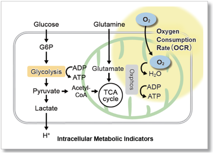
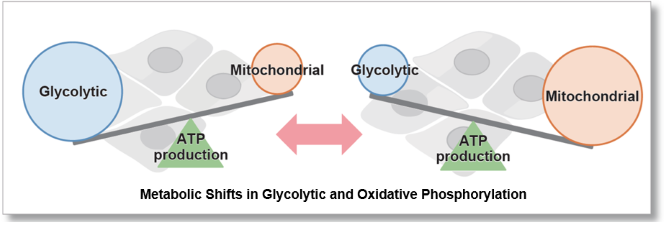
由于氧主要在线粒体氧化磷酸化产生三磷酸腺苷(ATP)的过程中消耗,因此其耗氧率(OCR)是分析线粒体功能的指标。众所周知,癌细胞通过糖酵解途径产生ATP,其效率低于氧化磷酸化。在免疫细胞中,氧化磷酸化的优势是抑制抗肿瘤,而糖酵解途径的优势促进抗肿瘤作用。因此,细胞的OCR作为能量代谢的检测指标。
图片1.png

图片2.png

产品概述

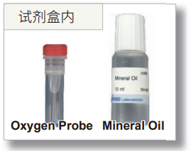
细胞外氧消耗量试剂盒包括氧气探针,其具有随着介质中氧气浓度的降低而增加荧光强度的特性,矿物油阻止氧气从空气中流入。

在用荧光酶标仪根据细胞外氧浓度测量荧光强度之后,根据Stern-Volmer方程计算细胞的OCR(自动计算表)。
1670202251340521.png1670202328366763.png

*该产品在群马大学Toshitada Yoshihara博士的指导下实现了产品化。

与现有方法比较

到目前为止,OCR测量需要昂贵的设备,如通量分析仪,实时动态检测酶标仪,以及酶标仪的功能调节。该试剂盒推荐给初此使用的人,因为它可以与常规荧光酶标仪一起使用,并附带所有必要试剂的完整包装。

image.png

与石英分析仪对比

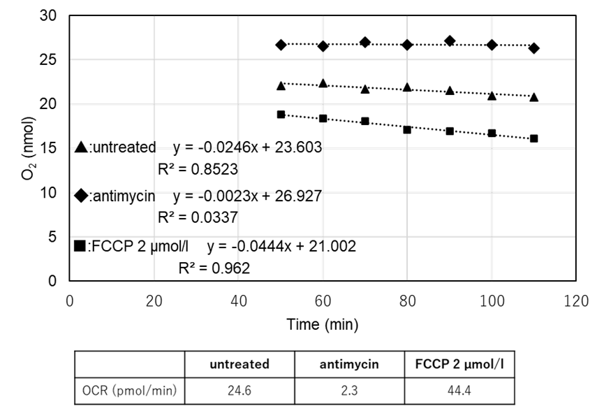
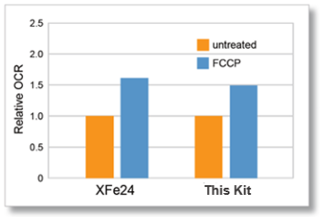
石英分析仪(XFe24)和本试剂盒在相同条件下(细胞类型、细胞数量和FCCP浓度)进行测量。

得到XFe24与本试剂盒相关氧消耗速度变化的数据。
图片6.png

细胞种类: HepG2

细胞数: 5×10⁴ cells/well

试剂: FCCP (Carbonyl cyanide 4-(trifluoromethoxy)phenylhydrazone)

FCCP 浓度: 2 μmol/l

实验例:抑制线粒体电子传输链

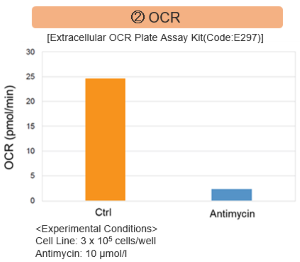
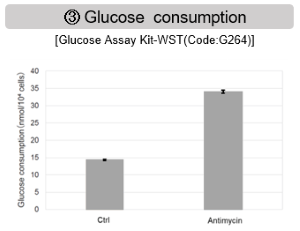
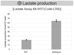
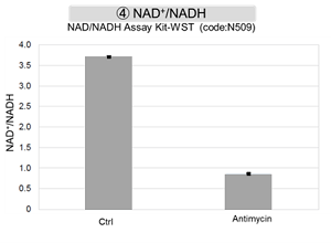
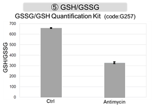
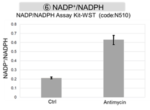
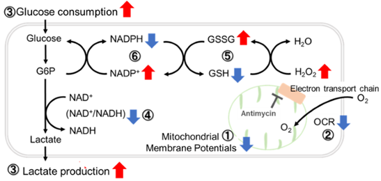
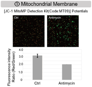
用抗霉素刺激大鼠细胞,评估线粒体电子运输链抑制后细胞状态的变化,检测多种指标。

结果表明,电子传输链的抑制导致(1)线粒体膜电位的降低和(2)OCR的降低。此外,观察到(3)整个糖酵解途径的NAD+/NADH比率降低,这是由于丙酮酸到乳酸的代谢增加,以维持糖酵解通路;(4)由于活性氧(ROS)增加,GSH耗竭;(6)由于谷胱甘肽生物合成所需NADPH减少,NADP+/NADPH比率增加。
图片10.png

1669944135247426.png图片12.png1669944181694051.png

图片14.png1669944219241446.png图片16.png

图片17.png

实验例:细胞最大呼吸能力评估

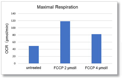
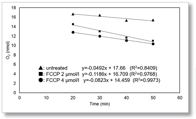
在HepG2细胞中,通过FCCP刺激后OCR值的变化来评估细胞的最大呼吸。

在FCCP浓度分别2µmol/l和4µmol/l 测量OCR。与2µmol/l相比,在4µmol/l时观察到OCR降低,表明在2µmol/l FCCP时最大呼吸。
图片9.png1669943932873010.png


细胞: HepG2

细胞数: 5×10⁴ cells/well

试剂: FCCP

FCCP 浓度 2, 4 μmol/l

实验例:不同细胞系代谢途径依赖性的比较

    许多癌症细胞通过糖酵解途径产生ATP。另一方面,最近有报道称,糖酵解途径被抑制的癌症细胞,可通过增强线粒体功能将能量代谢转移到OXPHOS而达到存活的目的,代谢途径的依赖性因细胞系不同而异。

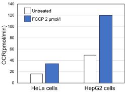
基于乳酸生成、ATP水平和OCR值,比较了两种癌症细胞HeLa和HepG2中OXPHOS和糖酵解的依赖性关系。

<通过乳酸生产和ATP水平进行评估>

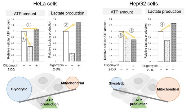
我们证实了当寡霉素刺激和糖酵解途径中的 2-Deoxy-D-glucose(2-DG)抑制OXPHOS的ATP合成时,ATP和乳酸产生的变化。结果表明,HeLa细胞依赖于糖酵解,HepG2细胞依赖于OXPHOS合成ATP。

*有关结果的更多信息,请参下方的“所用技术和产品的补充信息”部分。

所用技术和产品的补充信息
<通过乳酸产生和ATP的量进行评估>

当OXPHOS在HeLa细胞中被抑制时,ATP水平保持不变(①),乳酸产生增加(②)。这表明,即使OXPHOS被抑制,糖酵解也可以被进一步激活。相反,当糖酵解被抑制时,ATP水平显著降低(③),表明能量的产生依赖于糖酵解。另一方面,当OXPHOS在HepG2细胞中被抑制时,乳酸的产生增加(④),表明细胞试图通过增强糖酵解来补偿能量的产生,但ATP水平仍然降低(⑤)。这意味着,即使糖酵解增加,ATP的产生也没有得到足够的代偿。此外,当糖酵解被抑制时,ATP水平下降更多(⑥),这表明HepG2细胞的能量产生更多地依赖于OXPHOS而不是糖酵解。

5.jpg

本数据同时使用了:糖酵解/氧化磷酸化检测试剂盒—Glycolysis/OXPHOS Assay Kit(G270)

<OCR值评估>

使用相同数量的细胞,我们测量了当用线粒体解偶联剂FCCP刺激细胞来促进细胞耗氧量时的OCR值。结果表明,HepG2细胞比HeLa细胞具有更高的OCR值,这表明对OXPHOS的依赖性更强,这与ATP水平和乳酸产生的结果有关。
9.jpg

〈实验条件〉

细胞系:HeLa、HepG2

细胞数:5×10⁴个细胞/孔

刺激:FCCP

浓度:2μmol/l

使用:Oxygen Consumption Rate(OCR) Plate Assay Kit-氧消耗量检测试剂盒(货号:E297)进行评估

Q&A

Q:本试剂盒可以检测多少样本?
A:当测试一种细胞类型的相同数量的细胞时,可以测量24个样品。

*如果实验中使用了两种以上的细胞类型或多个细胞编号,则必须准备单独的空白和对照,并且可以测量的样本数量会有所不同。

有关详细信息,请参考手册中的板布局示例。

Q:悬浮细胞有什么实验案例吗?
A:我们准备了一个大鼠细胞实验的例子。<说明>

(1) 将大鼠细胞(3.0×10⁶细胞/ml)悬浮于RPMI培养基中作为空白3,将大鼠细胞(3.0×106细胞/ml)悬于工作溶液中作为对照或样品。将细胞接种在100µl(300000个细胞/孔)的96孔黑色透明底部微孔板中。


(2) 向空白1中加入100µl RPMI培养基,向空白2中加入100μl工作溶液。


(3) 将微孔板放置在预先设定为37°C的读板器中,孵育30分钟。


(4) 向空白1、空白2、空白3和对照品中加入10µl RPMI培养基。


(5) 将用RPMI培养基稀释的样品溶液(抗霉素或FCCP溶液)分10µl加入样品中。


(6) 加入样品溶液后,立即向每个孔中加入一滴矿物油。


(7) 将微板放置在37°C的平板读数器中,孵育5分钟。


(8) 在一个时间过程中,用荧光板读取器每10分钟测量一次强度,持续200分钟(Ex:500nm,Em:650nm,底部读数)。

(9) OCR值通过将获得的强度值输入下载的专用Excel计算表来计算。

每孔所需的样品和试剂数量。

image.png

1669944613174749.png

Q:如何使用此试剂盒计算OCR?
A:请使用Excel计算表并遵循以下说明


<OCR计算程序概述>

(1) 将OCR测量获得的强度值输入计算表,使用Stern-Volmer公式自动计算氧含量(nmol)。

(2) 根据时间(min)与氧含量(nmol)的关系图,检查所有测量条件下获得的线性范围。

(3) 计算步骤(2)中确认的时间(min)和氧含量(nmol)范围内的斜率。

(4) 根据步骤(3)中计算的斜率计算OCR(pmol/min)。

有关详细信息,请参阅手册中的“分析”。

*需要计算OCR的客户请至【网站首页】-【技术支持】-【实验工具】即可找到OCR计算器


Q:矿物油对细胞有细胞毒性吗?
A: 当通过Cell Counting Kit-8细胞毒性测定测定时,在用矿物油处理的细胞中未观察到毒性。


Q:为什么使用此试剂盒需要搭配可控温的酶标仪?
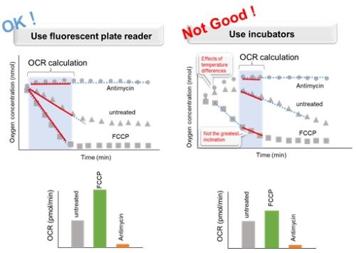
A: 在添加试剂和矿物油后,将微孔板与培养箱(或加热块、恒温室等)一起孵育,酶标仪中的温差将影响OCR结果。这导致数据再现性下降。因此,请使用温度可控的酶标仪。

<常规操作>

E297_2023_05.jpg

步骤3、7用于悬浮细胞,步骤5、9用于贴壁细胞

<孵育环境对结果的影响>

E297_2023_06.jpg

Q:OCR检测后如何测量细胞数
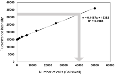
A:使用核酸探针(代码:H342)Hoechst 33342测量每个孔的细胞数,这是该方案的一个示例。

<说明>

(1) 将细胞接种到孔中进行OCR测量(液体体积:100μl/孔)。

(2) 将制备校准曲线的细胞接种到孔中(液体体积:100μl/孔)。

(3) OCR根据说明书进行测量。

(4) 向孔中加入10µl/孔的介质进行校准(使介质体积与OCR测量孔的体积对齐至110µl/孔)。

(5) 将用培养基稀释的Hoechst 33342溶液(10µg/ml)以100µl/孔的速度添加到所有孔中。

*从油的顶部添加OCR测量孔。

(6) 在37°C下培养30分钟。

(7) 用荧光板读数器(Ex:350nm,Em:461nm)测量。

(8) 制备校准曲线(X轴:细胞数量,Y轴:荧光强度),并计算用于OCR测量的孔中的细胞数量。

图片21.png

Q:可以长期存储工作液吗
A 工作液不能储存,需要现配现用。
Q:氧探针或矿物油的反复冷冻和解冻是否会影响测定?
A 我们已经证实,氧气探针和矿物油的反复冻融循环对测定没有影响。
Q:对照组与实验组之间OCR没有差异,有哪些可能得原因?
A请检查以下两个实验条件。

(1) 如果在测量过程中温度发生变化,可能会影响OCR结果。请确保以下两个步骤完全按照说明书执行。

・矿物油、溶剂和稀释溶剂等溶液在使用前应预热至37°C左右。

・加入试剂和矿物油后,请使用温度可控的酶标仪进行孵育。

请参阅Q&A“为什么使用此试剂盒需要搭配可控温的酶标仪?”。

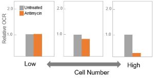
(2) 建议在最终计算前,优化单元格数据。如果细胞数量较低,实验组和对照组之间的差异也可能并不显著。

【带有细胞数和试剂处理的OCR值(预期结果图)】


参考文献


文献 研究对象 引用文献
1 细胞(HepG2) K.Saito.et al“Obesity-induced metabolic imbalance allosterically modulates CtBP2 to inhibit PPAR-alpha transcriptional activity”2023,Journal of Biological Chemistry,doi.org/10.1016/j.jbc.2023.104890
2 细胞(NIH3T3-L1) S. Oki, S. Kageyama, Y. Morioka and T. Namba, “Malonate induces the browning of white adipose tissue in high-fat diet induced obesity model”Biochem Biophys Res Commun.2023, doi:10.1016/j.bbrc.2023.08.054.
3 细胞

(Primary Hepatocyte)

S. Tsuno, K. Harada, M. Horikoshi, M. Mita, T. Kitaguchi, M. Y. Hirai, M. Matsumoto and T. Tsubo , ‘Mitochondrial ATP concentration decreases immediately after glucose administration to glucose-deprived hepatocytes’, FEBS Open Bio2023, doi:10.1002/2211-5463.13744.
  4 精子 “Arresting calcium-regulated sperm metabolic dynamics enables prolonged fertility in poultry liquid semen storage”, Scientific Reports 2023 , doi: 10.1038/s41598-023-48550-2.
5 细胞(HepG2) Takeo Nakanishi.et alAn implication of the mitochondrial carrier SLC25A3 as an oxidative stress modulator in NAFLDExperimental Cell Research.,2023,431,113740

Hampton蛋白结晶试剂盒Magnetic Base

上海金畔生物代理Hampton research品牌蛋白结晶试剂耗材工具等,我们将竭诚为您服务,欢迎访问Hampton research官网或者咨询我们获取更多相关Hampton research品牌产品信息。Products > Cryocrystallography > CrystalCap Systems > Magnetic Base

Magnetic Base

Features

  • Available in 2 strengths
  • Magnetic support for CrystalCap

Description

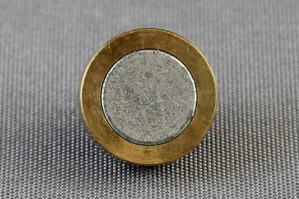
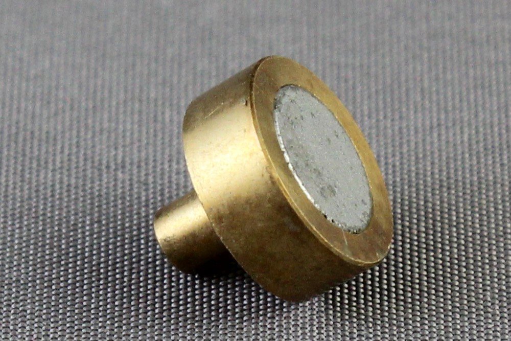
The Magnetic Base (9.3 mm diameter support) is a machined brass piece fitted with a magnet. The base fits into the goniometer head, providing a secure base for the CrystalCap System™ sample holders. Fits most goniometer heads. The Magnetic Base is available in two strengths: strong and medium. The stem of the magnet is 3 mm in diameter. The length of the stem is 5 mm. The thickness of the platform is 3.6 mm.


Magnetic Base

Click to Zoom In

Magnetic Base
Magnetic Base
Magnetic Base

CAT NO

HR4-943

NAME

Magnetic Base – Strong

DESCRIPTION

9,000 gauss – each

PRICE

$47.00

cart quote

CAT NO

HR4-627

NAME

Magnetic Base – Medium

DESCRIPTION

6,000 gauss – each

PRICE

$47.00

cart quote

Support Material(s)

Magnetic Base Magnetic Base Drawing

Related Item(S)

  • CrystalCap™
  • CrystalCap™ SPINE HT
  • CrystalCap™ ALS HT
  • CrystalCap™ ALS

Hampton蛋白结晶试剂盒Crystal Clear Sealing Tape

上海金畔生物代理Hampton research品牌蛋白结晶试剂耗材工具等,我们将竭诚为您服务,欢迎访问Hampton research官网或者咨询我们获取更多相关Hampton research品牌产品信息。Products > Crystallization Plates, Hardware & Accessories > Sealing Films, Tapes, Mats & Covers > Crystal Clear Sealing Tape

Crystal Clear Sealing Tape

Applications

  • Sealing tape used to seal sitting drop crystallization experiments

Features

  • Optically clear
  • Compatible with a wide range of crystallization reagents
  • UV compatible
  • Crystal Clear Mini Sealing Tape reseals and patches 96 well sitting drop plates
  • Crystal Clear Mini Sealing Tape seals Cryschem S and Cryschem M plate one row at a time

Description

These optically clear tapes are compatible with protein crystallization reagents. The polypropylene Crystal Clear Sealing Tapes use acrylic solvent based adhesives which are compatible with aqueous crystallization reagents.

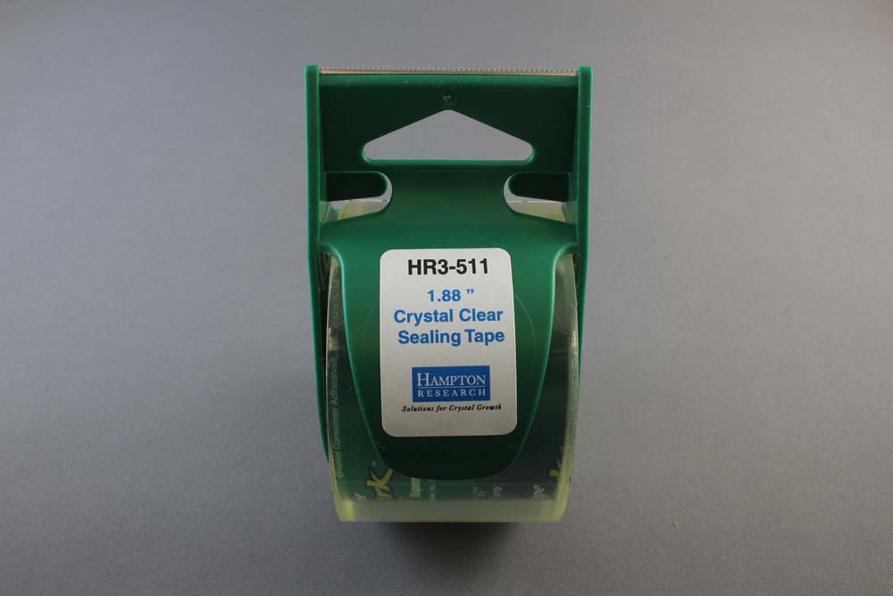
Catalog number HR3-511 is a 1.88 inch (48 mm) wide, 3 mil tape on a 43.7 yard (40 M) roll with a 1.5 inch core and is supplied with a green dispenser / cutter.

Catalog number HR4-511 is a 1.88 inch wide, 3 mil tape on a 109.3 yard roll with a 3 inch core and no dispenser / cutter.

Two strips of the HR3-511 or HR4-511 will seal a Corning, Cryschem, CrystalClear Strip, Greiner, Intelliplate, Linbro, Swissci (MRC), VDX and VDXm plate.

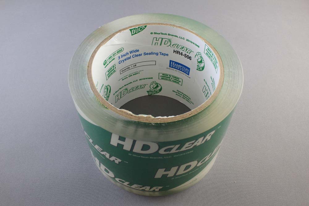
Catalog number HR4-506 is a 3 inch wide, 3 mil tape on a 54.86 yard roll with a 3 inch core and no dispenser / cutter.

One strip of the HR4-506 will seal a Corning, Cryschem M, CrystalClear Strip, Greiner, Intelliplate, Swissci (MRC) and VDXm plate.

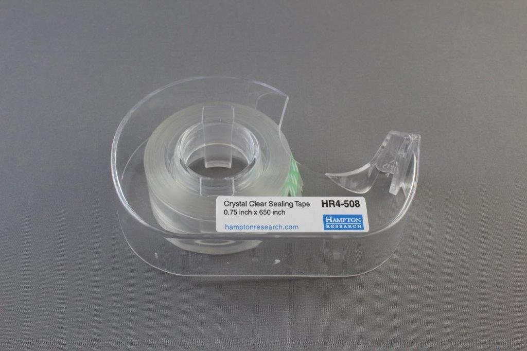
Crystal Clear Mini Sealing Tape HR4-508 is a 0.75 inch (19 mm) wide, 3 mil tape on a 650 inch (16.5 M) roll and is supplied with a clear dispenser / cutter. The tape is useful for resealing or patching wells after mounting crystals. The tape is also wide enough to seal a single row of a Cryschem S or Cryschem M plate.

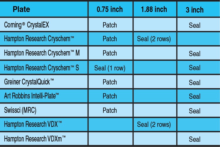
See chart for tape and plate compatibility.

Show More


Crystal Clear Sealing Tape

Click to Zoom In

Crystal Clear Sealing Tape

CAT NO

HR3-511

NAME

1.88 inch wide Crystal Clear Sealing Tape

DESCRIPTION

1.88 inch x 43.7 yard roll, with cutter

PRICE

$9.00

cart quote

CAT NO

HR4-511

NAME

1.88 inch wide Crystal Clear Sealing Tape

DESCRIPTION

1.88 inch x 60 yard roll

PRICE

$12.00

cart quote

CAT NO

HR4-506

NAME

3 inch wide Crystal Clear Sealing Tape

DESCRIPTION

3 inch x 55 yard roll

PRICE

$13.00

cart quote

CAT NO

HR4-508

NAME

0.75 inch wide Crystal Clear Mini Sealing Tape

DESCRIPTION

0.75 inch x 650 inch, with cutter

PRICE

$5.00

cart quote

Related Item(S)

  • X-Acto Gripster Knife

Hampton蛋白结晶试剂盒Seed Bead™ Kits

上海金畔生物代理Hampton research品牌蛋白结晶试剂耗材工具等,我们将竭诚为您服务,欢迎访问Hampton research官网或者咨询我们获取更多相关Hampton research品牌产品信息。Products > Tools & Seeding > Seed Bead > Seed Bead™ Kits

Seed Bead™ Kits

Applications

  • Generate seeds of protein crystals

Features

  • Easily generate consistent seed stocks
  • Use serial dilution to control the number of seeds introduced into the drop
  • Microseed matrix screening (MMS) seed stock
  • PTFE, Stainless Steel, Zirconium Silicate Ceramic, or Glass Seed Bead

Description

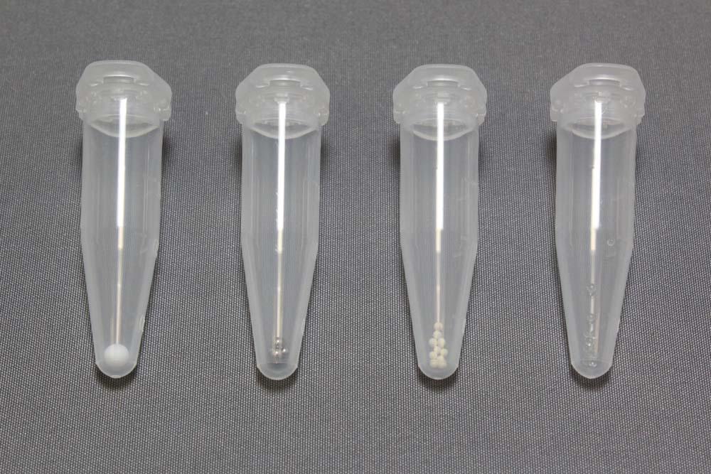
The Seed Bead kits are used to create a seed stock for performing subsequent seeding experiments. The Seed Bead kits contains 24 Seed Bead Tubes, a special 1.5 ml microcentrifuge tube, shaped to maintain the seed in the sample during mixing. Each tube is configured with a PTFE, Stainless Steel, Zirconium Silicate Ceramic, or Glass Seed Beads.

Seeding allows one to grow crystals in the metastable zone. Crystallization in this zone provides control, reproducibility and an improved likelihood of a successful crystallization experiment. Also, crystals can grow from seeds but cannot spontaneously nucleate. By placing a seed or solution of seeds in a drop which is saturated to the metastable zone, one can use the seeds to grow larger single crystals. By controlling the number of seeds introduced into the drop, one can control the number of crystals grown. It is not practically possible to measure and know the number of seeds introduced to a drop, but by performing serial dilutions from a concentrated seed stock, one can control the number of crystals grown in the drop.

Using the Seed Bead kits, one can create crystal seed stock for subsequent seeding experiments. Crystals are placed in the microcentrifuge tube with the Seed Bead(s) and either vortexed or sonicated to generate a seed stock. Then by performing serial dilutions, one can control the number and size of crystals in the experiment.

The Seed Bead kit is useful for the preparation of seed stocks for automated and semi-automated microseeding (D’Arcy 2007, Harlos 2008).

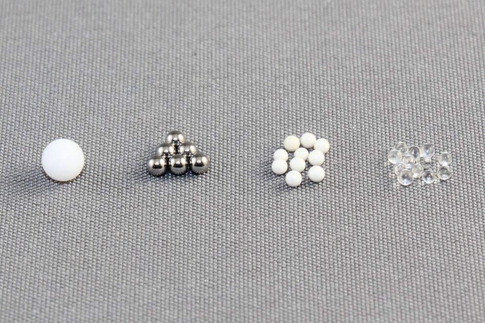
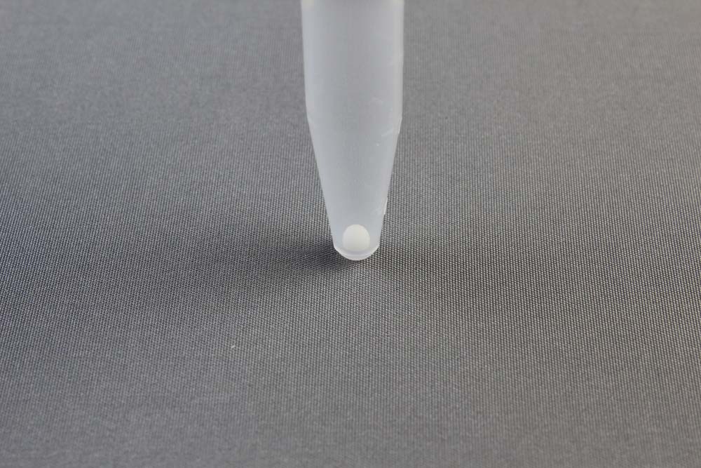
Each Seed Bead Kit (HR2-320) contains 24 special microcentrifuge tubes, each tube containing a single 3mm PTFE Seed Bead.

Each Seed Bead Steel Kit (HR4-780) contains 24 special microcentrifuge tubes, each tube containing a six 1.6 mm Stainless Steel Seed Beads.

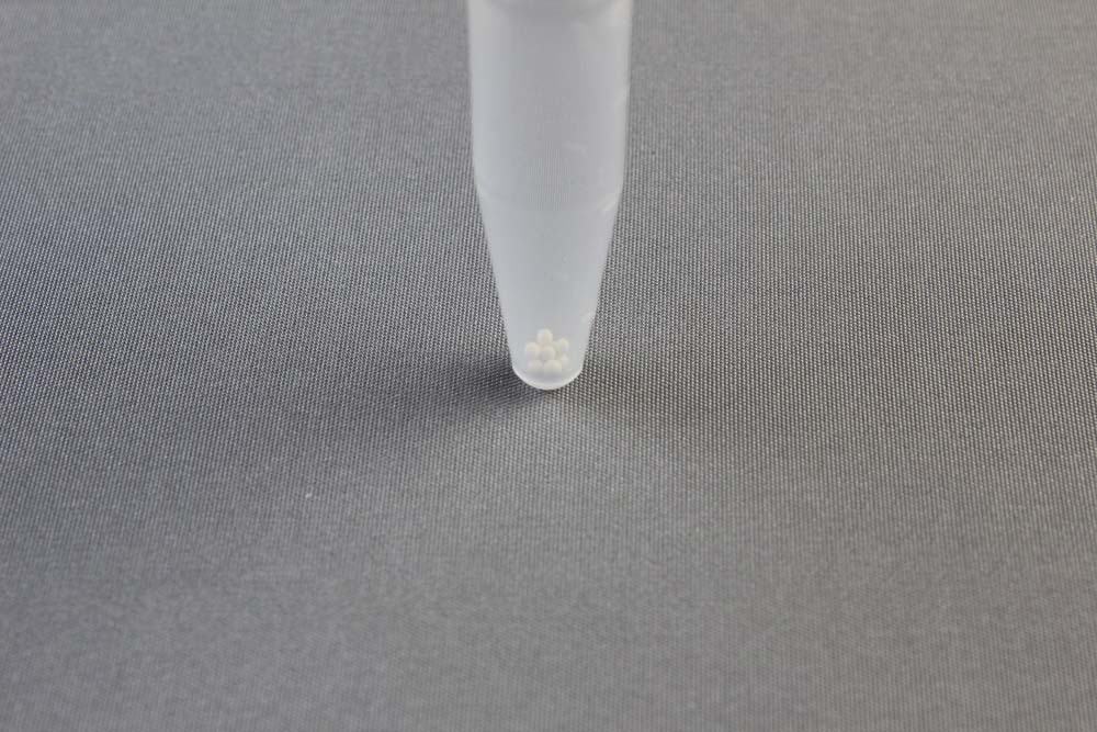
Each Seed Bead Ceramic Kit (HR4-781) contains 24 special microcentrifuge tubes, each tube containing a ten 1.0 mm Zirconium Silicate Ceramic Seed Beads.

Each Seed Bead Glass Kit (HR4-782) contains 24 special microcentrifuge tubes, each tube containing a ten 1.0 mm Glass Seed Beads.

Show More


Seed Bead™ Kits

Click to Zoom In

Seed Bead™ Kits
Seed Bead™ Kits
Seed Bead™ Kits
Seed Bead™ Kits
Seed Bead™ Kits
Seed Bead™ Kits

CAT NO

HR2-320

NAME

Seed Bead Kit

DESCRIPTION

24 tubes with PTFE Seed Bead

PRICE

$78.00

cart quote

CAT NO

HR4-780

NAME

Seed Bead Steel Kit

DESCRIPTION

24 tubes with Steel Seed Beads

PRICE

$75.00

cart quote

CAT NO

HR4-781

NAME

Seed Bead Ceramic Kit

DESCRIPTION

24 tubes with Ceramic Seed Beads

PRICE

$75.00

cart quote

CAT NO

HR4-782

NAME

Seed Bead Glass Kit

DESCRIPTION

24 tubes with Glass Seed Beads

PRICE

$75.00

cart quote

Support Material(s)

Seed Bead™ Kits HR2-320 Seed Bead User GuideSeed Bead™ Kits HR4-780 Seed Bead Steel User GuideSeed Bead™ Kits HR4-781 Seed Bead Ceramic User GuideSeed Bead™ Kits HR4-782 Seed Bead Glass User Guide

Related Item(S)

  • Crystal Crusher

References

1. Stura, E.A., Wilson, I.A., Methods: A Companion to Methods in Enzymology (1990) 1, 38-49.

2. Stura, E.A., Wilson, I.A., “Seeding Techniques” in Crystallization of Nucleic Acids and Proteins: A Practical Approach. Oxford University Press (1992) 99-126.

3. A method to produce microseed stock for use in the crystallization of biological macromolecules Luft, J.R., DeTitta, G.T. Acta Cryst. (1999). D55, 988-993.

4. J.R. Luft and G.T. DeTitta, Methods in Enzymology (1997) 276, 110-131.

5. Structure of an orthorhombic form of xylanase II from Trichoderma reesei and analysis of thermal displacement. Watanabe et al. Acta Cryst. (2006). D62, 784-792.

6. Crystallization and preliminary crystallographic analysis of p40phox, a regulatory subunit of NADPH oxidase. K. Honbou, S. Yuzawa, N. N. Suzuki, Y. Fujioka, H. Sumimoto and F. Inagaki. Acta Cryst. (2006). F62, 1021-1023 (Used seed bead to optimize)

7. Purification, crystallization and preliminary X-ray diffraction study of human ribosomal protein L10 core domain. Yuji Kobayashi et al. Acta Cryst. (2007). F63, 950-952

8. An automated microseed matrix-screening method. Allan D′Arcy, Frederic Villard and May Marsh. Acta Cryst. (2007). D63, 550-554

9. The role of bias in crystallization conditions in automated microseeding. Edwin Pozharski et al. Acta Cryst. (2008). D64, 1222-1227.

10. Transmission electron microscopy for the evaluation and optimization of crystal growth. Hilary P. Stevenson & Guillermo Calero et al. Acta Cryst. (2016) D72, 603-615.

11. Microseed matrix screening for optimization in protein crystallization: what have we learned? D’Arcy A, Bergfors T, Cowan-Jacob SW, Marsh M. Acta Crystallogr F Struct Biol Commun. 2014 Sep;70 (Pt 9):1117-26. doi: 10.1107/S2053230X14015507. Epub 2014 Aug 29.

Hampton蛋白结晶试剂盒X-Acto Gripster Knife

上海金畔生物代理Hampton research品牌蛋白结晶试剂耗材工具等,我们将竭诚为您服务,欢迎访问Hampton research官网或者咨询我们获取更多相关Hampton research品牌产品信息。Products > Tools & Seeding > X-Acto Gripster Knife > X-Acto Gripster Knife

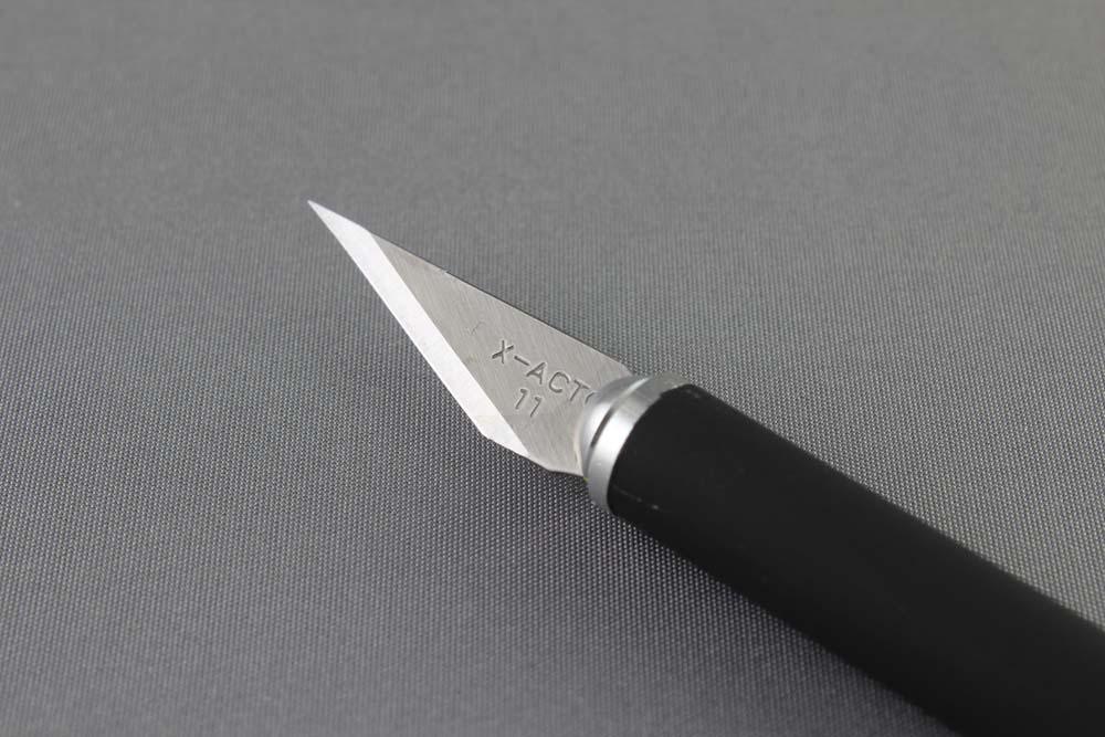
X-Acto Gripster Knife

Applications

  • Cutting and removing sealing tape and film from crystallization plates

Features

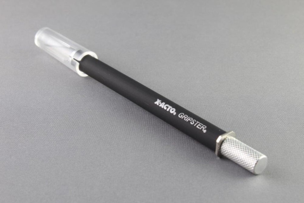
  • Sharp stainless steel blade
  • Safety cap for blade
  • Soft grip barrel
  • Anti-roll design – will not roll off table or microscope

Description

Precisely cuts tapes and films around reservoir and drops from crystallization plates, safely and easily. Ergonomic soft rubberized barrel is firm and comfortable for better control. Easy to clean handle. Rear blade release is for easier and safer blade changing. Rear blade release makes blade changing safer, easier, and faster. Safety cap cover for blade. Anti-roll stop design – knife will not roll off table or microscope and stab you in the toe. Supplied with one #11 blade. Replacement blades available separately.

#11 Replacement Blade – Extremely sharp point for fine angle cutting. Stainless steel. Sold in a package of five blades.

X-Acto® is a registered trademark of Elmer’s Products, Inc.

Show More


X-Acto Gripster Knife

Click to Zoom In

X-Acto Gripster Knife

CAT NO

HR4-124

NAME

X-Acto Gripster Knife

DESCRIPTION

Each

PRICE

$14.00

cart quote

CAT NO

HR4-126

NAME

Replacement #11 Blade, 5/pk

DESCRIPTION

5/pk

PRICE

$10.00

cart quote

Hampton蛋白结晶试剂盒LCP Sandwich Set

上海金畔生物代理Hampton research品牌蛋白结晶试剂耗材工具等,我们将竭诚为您服务,欢迎访问Hampton research官网或者咨询我们获取更多相关Hampton research品牌产品信息。Products > 96 Well Crystallization Plates > Paul Marienfeld > LCP Sandwich Set

LCP Sandwich Set

Applications

  • Crystallization screening of membrane proteins in lipidic mesophase as well as bicelle method and batch method

Features

  • Small volume LCP, bicelle and microbatch crystallization
  • High quality glass optics
  • Excellent drop optics since mesophase bolus is physically sandwiched between two optically clear surfaces eliminating the mesophase / aqueous medium interface and the corresponding roughness
  • Glass plate allows for visualization of microcrystals using birefringence-free examination between crossed polarizers
  • Superhydrophobic glass surfaces
  • Optimized for bright field, UV and fluorescent microscopy
  • Excellent stability, resistant to evaporation

Description

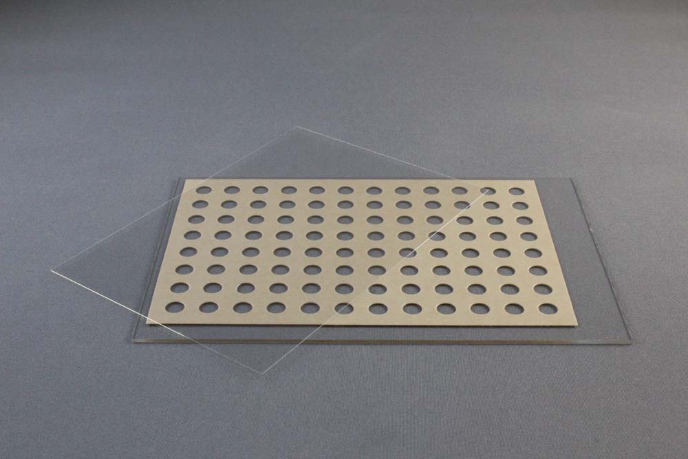
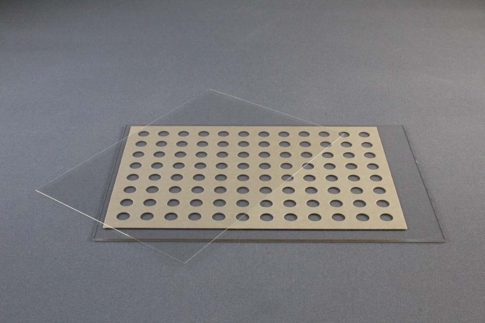
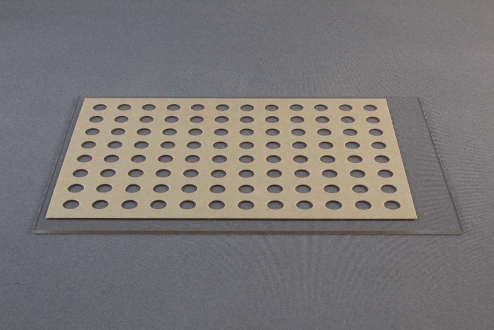
The LCP Sandwich Set consists of a base glass slide and an optimized cover slip. LCP Sandwich Set developed jointly with the renowned Scripps Research Institute in La Jolla, California, USA and is manufactured by Paul Marienfeld (0890003).

The LCP Glass Sandwich Set is a specially designed plate for either automated or manual setting of 96 Lipidic Cubic Phase matrix screening experiments. The plate can also be used for the bicelle method and batch method. The thin (< 2 mm high) plates have exquisite optical properties and are well suited for the detection of microcrystals and for birefringence-free imaging between crossed polarizers. The plates allow for in meso crystallization trials with 50 nanoliters protein/lipid mesophase and 1 microliter precipitant solution per trial.

The 96 well LCP Glass Sandwich Plate consists of a) 127.8 x 85.5 x 1 mm glass base plate with the footprint of an SBS-compliant plate, a 140 µm thick double sticky spacer with 96 punched out holes and b) a 0.2 mm thick glass coverslip. The double sticky spacer is already adhered to the base plate. A brown paper liner covers and protects the top of the double sticky spacer and base plate. The 0.2 mm thick glass coverslip fits over and seals the entire 96 well lower plate. Alternatively a series of twenty-four siliconized 18 x 18 mm No. 1 square cover slides (HR3-152) can be used to seal the entire 96 well lower plate. Each 18 x 18 mm No. 1 cover slide seals 4 wells. Space for a bar code is available at the left end of the plate. Each well can contain 50 nanoliters of cubic phase and 1 microliter of crystallization reagent.

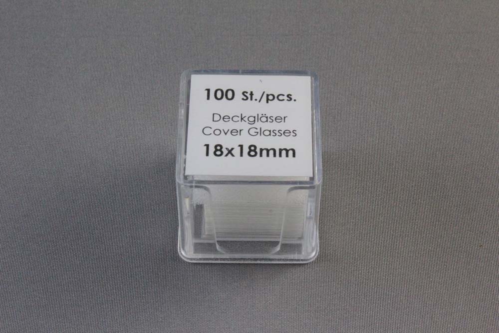
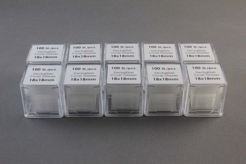
18 x 18 mm No. 1 Siliconized Cover Slides are made from chemically resistant borosilicate glass D 263 M of the first hydrolytic class. The slides are colorless, clear, and suitable for fluorescence microscopy. The slides feature a super hydrophobic surface on both sides. The slides are No. thickness (0.13 ro 0.16 mm). The slides are supplied in a two compartment plastic box, 100 slides per box, 10 boxes per case, 1,000 slides total for HR3-152.

The Tungsten-carbide glass cutter can be used for cutting and removal of the upper coverglass that seals the LCP Sandwich Set. Sold separately.

Show More


LCP Sandwich Set

Click to Zoom In

LCP Sandwich Set
LCP Sandwich Set
LCP Sandwich Set
LCP Sandwich Set
LCP Sandwich Set

CAT NO

HR3-151

NAME

LCP Sandwich Set

DESCRIPTION

Pack of 20

PRICE

$458.00

cart quote

CAT NO

HR3-152

NAME

18 x 18 mm No. 1 Siliconized Cover Slides, Squares

DESCRIPTION

Case of 10 packs

PRICE

$322.00

cart quote

Support Material(s)

LCP Sandwich Set HR3-151 LCP Sandwich Set User GuideLCP Sandwich Set HR3-151 LCP Sandwich Set Dimension Drawing

Related Item(S)

  • 4 Inch Soft Rubber Brayer
  • Tungsten Carbide Scribe & Glass Cutter with Replaceable Tips

References

1. Nano-volume plates with excellent optical properties for fast, inexpensive crystallization screening of membrane proteins. Vadim Cherezov and Martin Caffrey. J. Appl. Cryst. (2003). 36, 1372-1377.

2. A robotic system for crystallizing membrane and soluble proteins in lipidic mesophases. Vadim Cherezov, Avinash Peddi, Lalitha Muthusubramaniam, Yuan F. Zheng and Martin Caffrey. Acta Cryst. (2004). D60, 1795-1807 DOI: 10.1107/S0907444904019109.

3. Bicelle crystallization: a new method for crystallizing membrane proteins yields a monomeric bacteriorhodopsin structure. Faham, S. & Bowie, J. U. (2002). J. Mol. Biol. 316, 1-6.